MX1 / Myxovirus resistance protein 1 (HMV316)
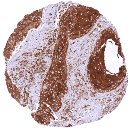
Recombinant Rabbit monoclonal / IgG 1:100 – 1:200 Research Use Only Intracellular Human HMV316 MX dynamin like GTPase 1,IFI-78K,IFI78,MX,MxA Prostate: An at least weak to moderate MX1 staining should be seen in basal cells while acinar cells remain negative. Colon: Crypt epithelial cells should be MX1 negative. MX1 is a GTP-binding protein with unknown biologic role. The interferon-induced GTP-binding protein MX1 ( myxovirus resistance protein 1) is coded by the MX1 gene located at 21q22.3. The gene consists of 17 exons and extends over 33 kb. MX1 is a member of a family of large GTPases which belongs to the dynamin superfamily. MX1 is a mediator of the resistance mechanisms to infections by influenza and other viruses both in cell culture and in transgenic mice. Its role in human influenza virus resilience is not fully understood, however, as humans harboring heterozygous and homozygous combinations of allelic MX1 variants could be linked to neither increased susceptibility to influenza virus nor to increased likelihood of severe disease. RNA expression data suggest that MX1 is ubiquitously expressed in normal tissues with a predominance in lymphatic tissues. A role in cancer biology has also been proposed for MX1. MX1 was found to be overexpressed in various cancer entities and the level of expression has been found to be linked to parameters of ... Images describing the MX1 staining pattern in normal tissues obtained by the antibody HMV316 are shown in our “ Normal Tissue Gallery ”. Brain Cerebrum MX1 staining of endothelial cells. Cerebellum MX1 staining of endothelial cells. Endocrine Tissues Thyroid Weak to moderate cytoplasmic MX1 staining of follicular cells. Parathyroid Negative. Adrenal gland Moderate cytoplasmic MX1 staining of medullary cells. Adrenocortical cells are MX1 negative or (focally) show weak positivity. Pituitary gland Negative. Respiratory system Respiratory epithelium Focal, weak to strong cytoplasmic MX1 staining in subsets of respiratory epithelial cells. Lung Pneumocytes are MX1 negative. Moderate to strong cytoplasmic MX1 staining of macrophages. Gastrointestinal Tract Salivary glands Moderate to strong cytoplasmic MX1 staining can be seen in some epithelial cell groups. Esophagus Weak to strong cytoplasmic MX1 staining of suprabasal cell layers. Stomach Weak to moderate cytoplasmic MX1 staining of surf... RNA data suggest that MX1 can be expressed in every tumor type. The clinical role is not clear yet – mostly because RNA data cannot discriminate the MX1 expression of tumor cells from stroma/inflammatory cells. The TCGA findings on MX1 RNA expression in different tumor categories have been summarized in the Human Protein Atlas. Gastric gastrointestinal stromal tumor (GIST)
with intense MX1 staining of tumor cells. MX1 negative basal cell carcinoma of the skin
showing a distinct MX1 staining of inflammatory
cells. Malignant mesothelioma of the pleura with
strong MX1 positivity of tumor cells. Cancer tissue gallery No data available at the moment IHC users have different preferences on how the stains should look like. Some prefer high staining intensity of the target stain and even accept some background. Others favor absolute specificity and lighter target stains. Factors that invariably lead to more intense staining include higher concentration of the antibody and visualization tools, longer incubation time, higher temperature during incubation, higher temperature and longer duration of the heat induced epitope retrieval (slide pretreatment). The impact of the pH during slide pretreatment has variable effects and depends on the antibody and the target protein. All images and data shown here and in our image galleries are obtained by the manual protocol described below. Other protocols resulting in equivalent staining are described as well. Manual protocol Freshly cut sections should be used (less than 10 days between cutting and staining). Heat-induced antigen retrieval for 5 minutes in an autoclave at 121°C in pH 7,8 Target ... The diagnostic and prognostic relevance of MX1 expression in tumors and in preneoplastic disease needs to be investigated. There are two ways how the specificity of antibodies can be documented for immunohistochemistry on formalin fixed tissues. These are: 1. Comparison with a second independent method for target expression measurement across a large number of different tissue types (orthogonal strategy), and 2. Comparison with one or several independent antibodies for the same target and showing that all positive staining results are also seen with other antibodies for the same target (independent antibody strategy). Orthogonal validation: For proteins such as MX1 which are expressed in virtually all tissues but restricted to specific cell types and cell compartments, orthogonal validation is not suited. For the antibody HMV316 specificity is, however, to some extent supported by the particularly high immunostaining level in lymphocytes and in cells of the hematopoesis. This is in agreement with RNA expression data from three independent RNA screening studies, including the Human Protein Atlas (HPA) RNA-s...