Myeloperoxidase / MPO (MSVA-692M)
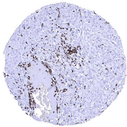
Mouse monoclonal / IgG 1:100 – 1:200 Research Use Only Lysozome Human MSVA-692M 4 kDa / 89 kDa myeloperoxidase; EC 1.11.1.7; EC1.11.2.2; fj80f04; MPO; mpx; myeloid-specific peroxidase; Myeloperoxidase; Myeloperoxidase heavy chain; Myeloperoxidase light chain Spleen: Numerous granulocytes with strong MPO staining should be seen within the red pulp of the spleen. Colon: MPO immunostaining should be absent in all epithelial cells of the colon while MPO positive granulocytes may be seen within capillaries or in the stroma of the lamina propria. MPO is a expressed in granulocytes and their precursor cells. Myeloperoxidase (MPO) is a 150 kDa peroxidase enzyme coded by the MPO gene on chromosome 17q23.1. MPO is most abundantly expressed in neutrophil granulocytes and stored in azurophilic granules. It comprises about 5% of the dry mass of neutrophil granulocytes. MPO is released to provide defense against invading pathogens. Its antimicrobial activity involves the production of acid. Antibodies against MPO which are also termed anti-neutrophil cytoplasmic antibodies (ANCAs) have been implicated in various types of vasculitis. MPO is considered a possible therapeutic target for coronary heart disease, inflammatory bowel disease and other conditions. A strong myeloperoxidase immunostaining is seen in neutrophil granulocytes while other granulocytes show a somewhat weaker and more variable staining. MPO positivity is also seen in granulocyte precursor cells in the bone marrow. MPO positive granulocytes occur in most normal tissues, either within blood vessels or also within the tissue. MPO positive material can be seen in the lumina of prostatic glands in some samples. In addition, a very weak cytoplasmic staining is seen in gastric glands which is considered a tolerable cross-reactivity. These findings are largely consistent with the RNA and protein data described in the Human Protein Atlas (Tissue expression MPO) Positive control: Spleen: Numerous granulocytes with strong MPO staining should be seen within the red pulp of the spleen. Negative control: Colon: MPO immunostaining should be absent in all epithelial cells of the colon while MPO positive granulocytes may be seen within capillaries or in the stroma of the lamina propria.... A variable content of MPO positive granulocytes is seen in cancers of various types. MPO expression by cancer cells is not seen. The TCGA findings on MPO RNA expression in different tumor categories have been summarized in the Human Protein Atlas. Esophageal squamous cell carcinoma containing numerous MPO positive granulocytes. Squamous cell carcinoma of the oral cavity containing numerous intravascular and stromal MPO positive granulocytes. Papillary renal cell carcinoma containing numerous MPO positive granulocytes. Cancer tissue gallery No data available at the moment IHC users have different preferences on how the stains should look like. Some prefer high staining intensity of the target stain and even accept some background. Others favor absolute specificity and lighter target stains. Factors that invariably lead to more intense staining include higher concentration of the antibody and visualization tools, longer incubation time, higher temperature during incubation, higher temperature and longer duration of the heat induced epitope retrieval (slide pretreatment). The impact of the pH during slide pretreatment has variable effects and depends on the antibody and the target protein. All images and data shown here and in our image galleries are obtained by the manual protocol described below. Other protocols resulting in equivalent staining are described as well. Manual protocol Freshly cut sections should be used (less than 10 days between cutting and staining). Heat-induced antigen retrieval for 5 minutes in an autoclave at 121°C in pH 7,8 Target ... Visualization and quantification of granulocytes for assessing the clinical role of these cells. The distinction of granulocytes in multicolor immunofluorescence is important in immune-oncological panels for the evaluation of the microenvironment of cancers. There are two ways how the specificity of antibodies can be documented for immunohistochemistry on formalin fixed tissues. These are: 1. comparison with a second independent method for target expression measurement across a large number of different tissue types (orthogonal strategy), and 2. Comparison with one or several independent antibodies for the same target and showing that all positive staining results are also seen with other antibodies for the same target (independent antibody strategy). Orthogonal validation is not completely suited for validation of granulocytes markers because these cells can be found in every organ. Findings obtained by MSVA-692M do, however, fit with data derived from three independent RNA screening studies, including the Human Protein Atlas (HPA) RNA-seq tissue dataset, the FANTOM5 project, and the Genotype-Tissue Expression (GTEx) project, which are all summarized in the Human Protein Atlas (Tissue expression MPO) . This is because the highest number o...