MYH7 / Myosin Heavy Chain 7 (MSVA-464M)
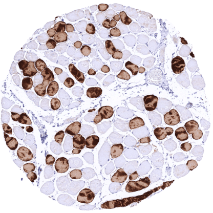
Mouse monoclonal / IgG2b, kappa 1:100 – 1:200 Research Use Only Cytoplasmic Human MSVA-464M Myosin-7, Myosin heavy chain 7, Myosin heavy chain slow isoform, Myosin heavy chain, cardiac muscle beta isoform, CMH1; MPD1; MyHC-beta; MyHC-slow; MYHCB; Myopathy, distal 1; Myosin heavy chain (AA 1-96); Myosin heavy chain slow isoform; Beta myosin heavy chain; cardiac muscle beta isoform; CMD1S; Myosin heavy chain, cardiac muscle beta isoform; Myosin, heavy chain 7, cardiac muscle, beta; Myosin, heavy polypeptide 7, cardiac muscle, beta; Myosin-7; Rhabdomyosarcoma antigen MU RMS 40.7A; SPMD; SPMM Skeletal muscle: A strong cytoplasmic MYH7 immunostaining should be seen in a subset of muscle cells. Skeletal muscle: A subset of skeletal muscle cells must completely lack MYH7 staining. MYH7 is a marker for type I fibers of skeletal muscle and of heart muscle. Myosin heavy chain 7 ( MYH7 ) is a 223,1 kDa protein coded by the MYH7 gene at chromosome 14q11.2. It represents a myosin heavy chain beta (MHC-β) (slow twitch) isoform expressed primarily in the heart where it defines the thick filament forming the cardiac muscle sarcomeres and plays a critical role in contraction. MYH7 is also built in the type I fibers of skeletal muscle. MYH7 is distinct in its enzymatic properties from the fast isoform of cardiac myosin heavy chain (MYH6, MHC-α). As compared to MYH 6, MYH7 has only about 25-50% the contractile velocity but 50% more actin attachment time. In the heart, MYH7 is predominantly expressed in the ventricle, while MYH6 is predominantly expressed in the atria. Multiple different mutations of MYH7 cause about 40% of all inherited (autosomal-dominant) hypertrophic cardiomyopathy (HCM) cases and are also responsible for paraspinal and proximal muscle atrophy. Images describing the MYH7 staining pattern in normal tissues obtained by the antibody MSVA-464M are shown in our “ Normal Tissue Gallery ”. Brain Cerebrum Negative. Cerebellum Negative. Endocrine Tissues Thyroid Negative. Parathyroid Negative. Adrenal gland Negative. Pituitary gland Negative. Respiratory system Respiratory epithelium Negative. Lung Negative. Gastrointestinal Tract Salivary glands Negative. Esophagus Negative. Stomach Negative. Duodenum Negative. Small intestine Negative. Appendix Negative. Colon Negative. Rectum Negative. Liver Negative. Gallbladder Negative. Pancreas Negative. Genitourinary Kidney Negative. Urothelium Negative. Male genital Prostate Negative. Seminal vesicles Negative. Testis Negative. Epididymis Negative. Female genital Breast Negative. Uterus, myometrium Negative. Uterus, ectocervix Negative. Uterus endocervix Negative. Uterus, endometrium Negative. Fallopian Tube Negative. Ovary Negative. Placenta early Negative. Placenta mature Negative. Amnion N... MYH7 immunostaining can be found in tumor cells with skeletal or heart muscle cell differentiation. The TCGA findings on MYH7 RNA expression in different tumor categories have been summarized in the Human Protein Atlas. MYH7 negative papillary renal cell carcinoma MYH7 negative squamous cell carcinoma showing infiltration of degenerated MYH7 positive skeletal muscle fibres MYH7 negative colorectal adenocarcinoma Cancer tissue gallery No data available at the moment IHC users have different preferences on how the stains should look like. Some prefer high staining intensity of the target stain and even accept some background. Others favor absolute specificity and lighter target stains. Factors that invariably lead to more intense staining include higher concentration of the antibody and visualization tools, longer incubation time, higher temperature during incubation, higher temperature and longer duration of the heat induced epitope retrieval (slide pretreatment). The impact of the pH during slide pretreatment has variable effects and depends on the antibody and the target protein. All images and data shown here and in our image galleries are obtained by the manual protocol described below. Other protocols resulting in equivalent staining are described as well. Manual protocol Freshly cut sections should be used (less than 10 days between cutting and staining). Heat-induced antigen retrieval for 5 minutes in an autoclave at 121°C in pH 7,8 Target ... MYH7 can be used for studying the role of different skeletal muscle fibres because MYH7 allows a distinction of slow (MYH7 positive) from fast (MYH7 negative) isoform of cardiac myosin heavy chain. MYH7 has attracted considerable interest as a result of its fundamental functions in cardiac and skeletal muscle contraction In principle, there are two ways how the specificity of antibodies can be documented for immunohistochemistry on formalin fixed tissues. These are: 1. Comparison with a second independent method for target expression measurement across a large number of different tissue types (orthogonal strategy), and 2. Comparison with one or several independent antibodies for the same target and showing that all positive staining results are also seen with other antibodies for the same target (independent antibody strategy). Orthogonal validation: For the antibody MSVA-464M specificity is suggested by the full concordance of the immunostaining data with data from three independent RNA screening studies, including the Human Protein Atlas (HPA) RNA-seq tissue dataset, the FANTOM5 project, and the Genotype-Tissue Expression (GTEx) project, which are all summarized in the Human Protein Atlas (Tissue expression MYH7) . RNA expression was only described in these organs where a positive MYH7 immunostaining...