Napsin A (MSVA-112R)
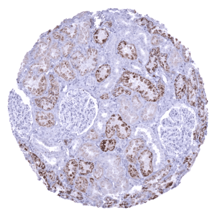
Recombinant Rabbit monoclonal / IgG 1:100 – 1:200 Research Use Only Cytoplasmic Human MSVA-112R ASP4, Aspartyl protease 4, KAP, Kidney derived aspartic protease like protein (Kdap), NAP1, NAPA, Napsa, napsin A aspartic peptidase, Pronapsin A, SNAPA Kidney: an at least moderate granular cytoplasmic staining should be seen in proximal tubule cells Colon: staining should be absent in epithelial and other cells of the lamina propria. Novel aspartic proteinase of the pepsin family A (Napsin A, TAO1/TAO2) belongs to the peptidase A1 family, together with Cathepsin E, renin, and pepsin. Napsin A is a proteinase, that cleaves proteins and peptides to produce mature or active forms of these molecules. Napsin A is known to be expressed in type II pneumocytes, intra-alveolar macrophages, and in renal tubules. Physiologically, Napsin A is involved in the maturation of prosurfactant protein B in type II pneumocytes, phagocytosis by macrophages,and protein catabolism in renal cells. In the lung, Napsin A is regulated by the thyroid transcription factor 1 (TTF1). Napsin A expression is limited to few cancer types. Napsin A immunohistochemistry is therefore highly useful for distinction of different tumor types in the lung and in the ovary as well as for assessing tumors of unknown origin.[1] Novel aspartic proteinase of the pepsin family A (Napsin A, TAO1/TAO2) belongs to the peptidase A1 family, together with Cathepsin E, renin, and pepsin. Napsin A is a proteinase, that cleaves proteins and peptides to produce mature or active forms of these molecules. Napsin A is known to be expressed in type II pneumocytes, intra-alveolar macrophages, and in renal tubules. Physiologically, Napsin A is involved in the maturation of prosurfactant protein B in type II pneumocytes, phagocytosis by macrophages , and protein catabolism in renal cells. In the lung, Napsin A is regulated by the thyroid transcription factor 1 (TTF1). Napsin A expression is limited to few cancer types. Napsin A immunohistochemistry is therefore highly useful for distinction of different tumor types in the lung and in the ovary as well as for assessing tumors of unknown origin. [1] [1] Weidemann et al. “Napsin A Expression in Human Tumors and Normal Tissues” Pathology & Oncology Research 2021; 27: 613099 Napsin staining pattern in Normal Tissues with antibody MSVA-112R (images are shown in our “Normal Tissue Gallery”) Brain Cerebrum Negative. Cerebellum Negative. Endocrine Tissues Thyroid Negative. Parathyroid Negative. Adrenal gland Negative. Pituitary gland Negative. Respiratory system Respiratory epithelium Negative. Lung Very strong cytoplasmic Napsin A staining in type II pneumocytes and in alveolar macrophages. Gastrointestinal Tract Salivary glands Negative. Esophagus Negative. Stomach Negative. Colon Negative. Duodenum Negative. Rectum Negative. Small intestine Negative. Liver Negative. Gallbladder Negative. Pancreas Negative. Genitourinary Kidney Napsin A staining is variable and ranges from weak to strong. Proximal tubuli stain more strongly than distal tubuli. Some collecting ducts also stain positive. Urothelium Negative. Male genital Prostate Negative. Seminal vesicles Negative. Testis Negative. Epididymis Moderate to strong Napsin A positivity occurs at the luminal pole o... The most commonly Napsin A positive cancer types include: adenocarcinoma of the lung (approx. 85% positive), clear cell adenocarcinoma of the ovary (70%) and the endometrium (40%), papillary renal cell carcinoma (40%), small cell carcinoma of the lung (20%), endometroid serous carcinoma (10%), papillary thyroid carcinoma (10%) and clear cell renal cell carcinoma (10%). The TCGA findings on Napsin A RNA expression in different tumor categories have been summarized in the Human Protein Atlas. Strong Napsin A positivity in an adenocarcinoma of the lung. Napsin A negative squamous cell carcinoma of the lung. Strong Napsin A immunostaining of remnant non-neoplastic lung tissue and/or alveolar macrophages. Clear cell renal cell carcinoma without any Napsin A immunostaining. Cancer tissue gallery Napsin A (MSVA-112R) publication summary Relevant publication: Weidemann et al. “Napsin A Expression in Human Tumors and Normal Tissues” Published in Pathology & Oncology Research 2021; 27: 613099 A total of 11957 tumors were analyzed from 115 different tumor categories by using the following protocol: Heat-induced antigen retrieval for 5 minutes in an autoclave at 121°C in pH7,8 Target Retrieval Solution buffer. MSVA-112R at a dilution of 1:200 at 37°C for 60 minutes. Visualization of bound antibody by the EnVision Kit (Dako, Agilent). This protocol was also used for all stainings depicted in our tumor and normal tissue galleries. At least one case with a positive Napsin A immunostaining was seen in 16 (13,9%) and at least one case with a strong Napsin A immunostaining was seen in 6 (5,2%) of 115 tumor categories. The distribution of positive staining results is shown “organ-systematic” and in a “ranking order” figure below (images based on data from Weidemann et al ). Results on p... IHC users have different preferences on how the stains should look like. Some prefer high staining intensity of the target stain and even accept some background. Others favor absolute specificity and lighter target stains. Factors that invariably lead to more intense staining include higher concentration of the antibody and visualization tools, longer incubation time, higher temperature during incubation, higher temperature and longer duration of the heat induced epitope retrieval (slide pretreatment). The impact of the pH during slide pretreatment has variable effects and depends on the antibody and the target protein. All images and data shown here and in our image galleries are obtained by the manual protocol described below. Other protocols resulting in equivalent staining are described as well. Manual protocol Freshly cut sections should be used (less than 10 days between cutting and staining). Heat-induced antigen retrieval for 5 minutes in an autoclave at 121°C in pH 7,8 Target ... A comprehensive study analyzing Napsin-A expression in various different tumor entities would be helpful to assess the diagnostic significance of Napsin-A IHC. The clinical role of Napsin A expression levels in lung or renal cancers is unknown. Specificity of MSVA-112R is documented by strong positive staining in cell types that are well documented to express Napsin A such as pneumocytes and alveolar macrophages in the lung, proximal tubuli in the kidney and epithelial cells in the epididymis. In addition Napsina A immunostaining was absent in all tissues known to not express Napsin A including tissues notorious for non-specific IHC background such as kidney, colonic mucosa, and epidermis. Normal tissue gallery