Occludin (MSVA-415M)
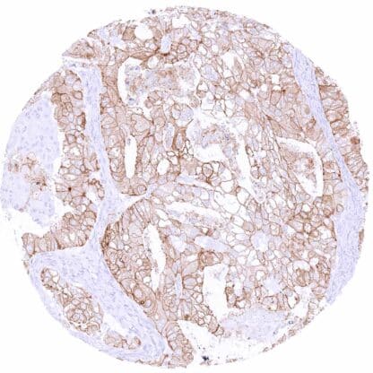
Mouse monoclonal / IgG 1:100 – 1:200 Research Use Only Cell Surface and Cytoplasmic Human MSVA-415M BLCPMG; Occludin; OCLN; Phosphatase 1 regulatory subunit 115; PPP1R115; PTORCH1; Tight junction protein occludin Colon: A strong membranous occludin staining of epithelial cells should be seen with a particularly strong staining at the apical/luminal membranes. Colon: All non-epithelial and non-endothelial cells must not show any occludin staining. Occludin is a Pivotal component of tight junctions. Occludin ( OCLN) is a 65 kDa plasma-membrane protein coded by the OCLN gene at chromosome 5q13.1. The Occludin protein is preferentially located at the tight junctions of epithelial and endothelial cells. Together with other proteins such as claudins and zonula occludens-1 (ZO-1), occludin regulates the formation, maintenance, and function of tight junctions. In particular, occludin may be important for tight junction stability and barrier function. As a NADH oxidase, occludin influences critical aspects of cell metabolism like glucose uptake, ATP production and gene expression. Manipulation of the occludin content in human cells affects the expression of glucose transporters, and the activation of transcription factors such as NFkB. In cancer, loss of occludin has been linked to increased invasion, reduced adhesion and metastasis. Occludin staining pattern in Normal Tissues with antibody MSVA-415M (images are shown in our “ Normal Tissue Gallery ”) Brain Cerebrum Negative. Cerebellum Negative. Endocrine Tissues Thyroid Intense membranous occludin immunostaining of follicular cells. Parathyroid Moderate membranous occludin staining of epithelial cells. Adrenal gland Negative. Pituitary gland Only few epithelial cells show a detectable membranous occluding positivity in the adenohypophysis. The neurohypophysis is occludin negative. Respiratory system Respiratory epithelium Significant membranous occludin staining. Lung Both pneumocytes and endothelial cells show a significant membranous occludin staining while macrophages remain negative. Gastrointestinal Tract Salivary glands Membranous occludin staining is most intense in excretory ducts and only faint in glandular cells. Esophagus Occludin staining predominates in intermediate cell layers of the squamous epithelium, while occludin positivity is either absent or... At variable levels, occludin immunostaining occurs in many different tumor entities. However, as compared to normal tissues, occludin expression may often be lower. Reduced expression of occludin in cancer has been linked to poor prognosis in studies. The TCGA findings on Occludin RNA expression in different tumor categories have been summarized in the Human Protein Atlas. Medullary cancer with moderate to strong occludin staining of tumor cells Adenocarcinoma (Gleason 5+5=10) with intense membranous and cytoplasmic occludin staining of tumor cells Adenocarcinoma with strong occludin staining of most tumor cells Cancer tissue gallery No data available at the moment IHC users have different preferences on how the stains should look like. Some prefer high staining intensity of the target stain and even accept some background. Others favor absolute specificity and lighter target stains. Factors that invariably lead to more intense staining include higher concentration of the antibody and visualization tools, longer incubation time, higher temperature during incubation, higher temperature and longer duration of the heat induced epitope retrieval (slide pretreatment). The impact of the pH during slide pretreatment has variable effects and depends on the antibody and the target protein. All images and data shown here and in our image galleries are obtained by the manual protocol described below. Other protocols resulting in equivalent staining are described as well. Manual protocol Freshly cut sections should be used (less than 10 days between cutting and staining). Heat-induced antigen retrieval for 5 minutes in an autoclave at 121°C in pH 7,8 Target ... The prognostic impact of occludin expression levels are unclear for many tumor types. Studies comparing occludin expression levels between different tumor entities are lacking. There are two ways how the specificity of antibodies can be documented for immunohistochemistry on formalin fixed tissues. These are: 1. comparison with a second independent method for target expression measurement across a large number of different tissue types (orthogonal strategy), and 2. Comparison with one or several independent antibodies for the same target and showing that all positive staining results are also seen with other antibodies for the same target (independent antibody strategy). Orthogonal validation: Orthogonal validation is not applicable for occludin antibodies because of the ubiquitous expression of the protein in blood vessels and epithelial cells in virtually all organs. The conspicuous absence of MSVA-415M staining seen in adrenal gland, muscle, and bone marrow as well as the low staining in prostate and the adenohypophysis is, however, consistent with data from three independent RNA screening studies, including the Human Protein Atlas (HPA) RNA-seq tissue dat...