p63 (MSVA-063R)
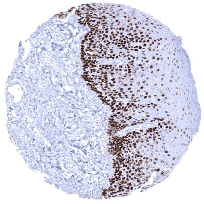
Recombinant Rabbit monoclonal / IgG 1:100 – 1:200 Research Use Only Nuclear Human MSVA-063R Amplified in squamous cell carcinoma (AIS); Chronic ulcerative stomatitis protein (CUSP); EEC3; Keratinocyte transcription factor KET; LMS; NBP; p40; P51/P63; p53 like transcription factor; p53-related protein p63; RHS; SHFM4; TAp63alpha; TP53CP; TP53L; TP63; TP73; TP73L; Transformation-related protein 63; Trp53rp1; Trp6;3; Tumor protein 63; Tumor protein p53-like; tumor protein p73-like Tonsil: Virtually all squamous epithelial cells must show a moderate to strong, nuclear staining, while few scattered lymphocytes and endothelial cells must show a at least a weak staining. Tonsil: The vast majority of lymphocytes should be p63 negative. p63 is expressed in cells with squamous, urothelial, or myoepithelial differentiation. Tumor protein 63 (p63) is a transcription factor of the p53 gene family encoded by the TP63 gene located at chromosome 3q28. Together with p73 it makes up the p53 gene family based on their structural similarity. p63 regulates the activity of a multitude of genes involved in growth and development of the ectoderm and derived structures and tissues, such as basal layer keratins and cell cycle control genes. Accordingly, p63 expression is found in basal cell layers of various organs, squamous epithelial cells of many organs and urothelium. p63 (syn. TAp63) is closely related to p40 (syn. ΔNp63) as both proteins represent isoforms of the p63 gene with distinct molecular functions. While “full length” p63 (TAp63) activates p53 target genes such as p21 or BAX, the shorter transcript p40 (ΔNp63) inhibits activation of p53 and “full length” p63. p63 -/- mice suffer from severe developmental defects which include the lack of limbs, teeth, and mammary glands. Brain Cerebrum Negative. Cerebellum Negative. Endocrine Tissues Thyroid Negative. Parathyroid Negative. Adrenal gland Negative. Pituitary gland Negative. Respiratory system Respiratory epithelium Strong nuclear p63 positivity of basal cells. Lung Negative. Gastrointestinal Tract Salivary glands Strong nuclear p63 positivity of myoepithelial cells. Esophagus Nuclear p63 staining in squamous epithelial cells is strongest in the basal cell layers while the intensity gradually decreases towards the top layers. Stomach Negative. Duodenum Negative. Small intestine Negative. Appendix Negative. Colon Negative. Rectum Negative. Liver Negative. Gallbladder Negative. Pancreas Negative. Genitourinary Kidney Negative. Urothelium Strong nuclear p63 staining in all urothelial cells except umbrella cells. Male genital Prostate Strong nuclear p63 positivity of basal cells. Seminal vesicles Strong nuclear p63 positivity of basal cells. Testis Negative. Epididymis Strong nuclear p63 positivity of basal c... p63 immunostaining is predominantly seen in cancers that are derived from p63 positive normal cell types. The most commonly positive cancers include squamous cell carcinomas of all origins, urothelial carcinomas, thymic tumors, basal cell carcinomas, and various salivary gland tumors. p63 can also be expressed in a small fraction of tumors from entities that are derived from p63 negative normal tissues. In some of these tumors, p63 neo-expression is linked to focal squamous cell differentiation which can for example occur in endometroid cancer and malignant mixed Mullerian tumors of the uterus, ovarian, pancreatic and cholangiocellular carcinomas. In other tumors, occasional p63 positive cells may reflect stemness properties. The TCGA findings on p63 RNA expression in different tumor categories have been summarized in the Human Protein Atlas. Urinary bladder- Non-invasive urothelial carcinoma (low grade, pTaG2) showing strong p63 positivity of tumor cells Colon- p63 negative colorectal... No data available at the moment IHC users have different preferences on how the stains should look like. Some prefer high staining intensity of the target stain and even accept some background. Others favor absolute specificity and lighter target stains. Factors that invariably lead to more intense staining include higher concentration of the antibody and visualization tools, longer incubation time, higher temperature during incubation, higher temperature and longer duration of the heat induced epitope retrieval (slide pretreatment). The impact of the pH during slide pretreatment has variable effects and depends on the antibody and the target protein. All images and data shown here and in our image galleries are obtained by the manual protocol described below. Other protocols resulting in equivalent staining are described as well. Manual protocol Freshly cut sections should be used (less than 10 days between cutting and staining). Heat-induced antigen retrieval for 5 minutes in an autoclave at 121°C in pH 7,8 Target ... A comprehensive study analyzing p63 expression in various different tumor entities would be helpful to assess the diagnostic significance of p63 IHC. The roles of p63 in multiple aspects of cancer, including tumorigenesis, cancer progression, and metastasis as well as how they impact other diseases are still not completely discovered. There are two ways how the specificity of antibodies can be documented for immunohistochemistry on formalin fixed tissues. These are: 1. Comparison with a second independent method for target expression measurement across a large number of different tissue types ( orthogonal strategy ), and 2. Comparison with one or several independent antibodies for the same target and showing that all positive staining results are also seen with other antibodies for the same target ( independent antibody strategy ). Orthogonal validation: For the antibody MSVA-063R , specificity is suggested by the perfect concordance of the immunostaining data with data from three independent RNA screening studies, including the Human Protein Atlas (HPA) RNA-seq tissue dataset, the FANTOM5 project, and the Genotype-Tissue Expression (GTEx) project, which are all summarized in the Human Protein Atlas (Tissue expression p63) . P63 RNA expression was only seen in organs covered by squamous epithelium (esophagus, vagina...