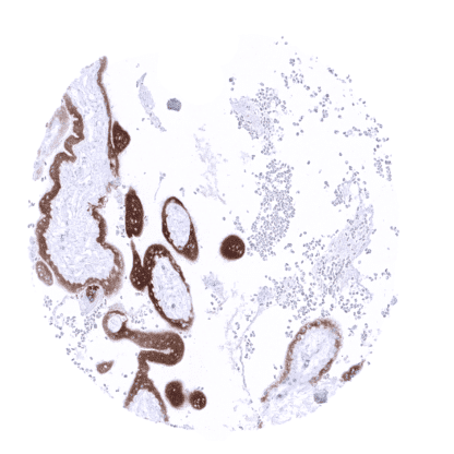
PAPP-A (MSVA-780M)
Mouse monoclonal / IgG 1:100 Research Use Only Cytoplasmic. Human MSVA-780M ASBABP2; Aspecific BCL2 ARE binding protein 2; Differentially placenta 1 expressed protein; DIPLA1; IGF-dependent; IGFBP4ase; Insulin-like growth factor-dependent IGF-binding protein 4 protease (IGFBP-4 protease); PAPA; PAPP A; PAPPA1; Pappalysin-1; Pregnancy Associated Plasma Protein A (PAPP-A) Placenta:Trophoplast and chorion cells should show a strong cytoplasmic papp-A staining. Colon:Papp-A immunostaining should be completely absent in both stromal end epithelial cells. PAPP-A is expressed in the placenta. Pregnancy-associated plasma protein A (Papp-A) is a metalloproteinase encoded by the PAPPA gene located at chromosome 9q33.2. The protein is secreted, activated upon collagen binding, and is thought to be involved in local proliferative processes such as wound healing and bone remodeling. Its main substrates are insulin-like growth factor binding proteins which are cleaved by papp-A. Papp-A is mainly produced in the placenta and has been connected with paracrine and autocrine control of trophoblast invasion of the decidua. Low maternal serum papp-A concentrations have been implicated with preeclampsia and Down syndrome. Papp-A is a placenta specific protein which is seen both in the early and mature placenta. Strongest immunostaining is seen in syncytiotrophoblast and in chorion cells. Staining may be less or even absent in the cytotrophoblast. These findings are largely comparable to the RNA and protein data described in the Human Protein Atlas (Tissue expression PAPP-A) Positive control: Placenta: Trophoplast and chorion cells should show a strong cytoplasmic papp-A staining. Negative control: Colon: Papp-A immunostaining should be completely absent in both stromal end epithelial cells. In the placenta, chorion cells show a moderate to strong papp-A immunostaining. Staining is also seen in the stroma, probably due to the „secreted“ nature of the protein. Trophoblast cells of a first trimenon placenta exhibiting a moderate to strong papp-A immunostaining. Papp-A immunostaining is absent in colon mucosa. Normal tissue gallery Papp-A is not known to be expressed in cancers. The TCGA findings on PAPP-A RNA expression in different tumor categories have been summarized in the Human Protein Atlas. Papp-A negative invasive breast cancer of no special type (NST). Papp-A negative clear cell renal cell carcinoma. Papp-A negative gastrointestinal stromal tumor (GIST) of the stomach. Cancer tissue gallery No data available at the moment IHC users have different preferences on how the stains should look like. Some prefer high staining intensity of the target stain and even accept some background. Others favor absolute specificity and lighter target stains. Factors that invariably lead to more intense staining include higher concentration of the antibody and visualization tools, longer incubation time, higher temperature during incubation, higher temperature and longer duration of the heat induced epitope retrieval (slide pretreatment). The impact of the pH during slide pretreatment has variable effects and depends on the antibody and the target protein. All images and data shown here and in our image galleries are obtained by the manual protocol described below. Other protocols resulting in equivalent staining are described as well. Manual protocol Freshly cut sections should be used (less than 10 days between cutting and staining). Heat-induced antigen retrieval for 5 minutes in an autoclave at 121°C in pH 7,8 Target ... It is currently unknown, whether Papp-A expression can occur in cancer. There are two ways how the specificity of antibodies can be documented for immunohistochemistry on formalin fixed tissues. These are: 1. comparison with a second independent method for target expression measurement across a large number of different tissue types (orthogonal strategy), and 2. Comparison with one or several independent antibodies for the same target and showing that all positive staining results are also seen with other antibodies for the same target (independent antibody strategy). Orthogonal validation: For the antibody MSVA-780M specificity is suggested by the full concordance of the immunostaining with RNA expression data derived from the Human Protein Atlas (HPA) RNA-seq tissue dataset , the FANTOM5 project, and the Genotype-Tissue Expression (GTEx) project which are all summarized in the protein atlas. Immunostaining by using MSVA-780M was exclusively detected in the placenta, the only organ with documented RNA expression. Comparison of antibodies: True expression ...