PAX8 (MSVA-708R)
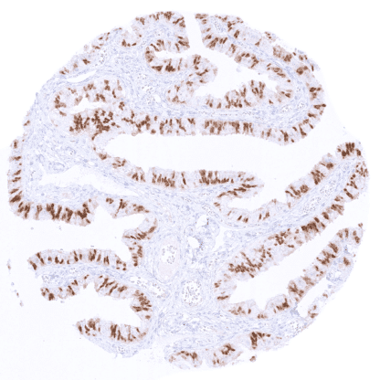
Recombinant Rabbit monoclonal / IgG 1:100 – 1:200 Research Use Only Nuclear Human MSVA-708R Paired box gene 8; Paired domain gene 8; PAX8; paired box homeotic gene 8 Kidney: At least weak to moderate, distinct nuclear staining should be seen in the majority of epithelial cells of the proximal and distal renal tubules, collection ducts, and the parietal epithelial cells of the Bowman’s capsule. Tonsil: No staining should be seen in squamous epithelial cells and in lymphocytes. PAX8 is a marker for kidney, thyroid and Mueller tract tissues. PAX8 ( Paired box gene 8) is a member of the paired-box gene family. The 48kDa protein is coded by the PAX8 gene at chromosome 2q14.1. PAX8 is physiologically expressed in organ development of the thyroid, Wollffian and Muellerian tract, renal/upper urinary tract, and the inner ear, and is also required for tissue homeostasis in the respective adult tissue. PAX8 is a master transcriptional regulator of thyroid specific genes such as thyroglobulin, thyroid peroxidase, and the sodium-iodide symporter by binding to the promoter regions. In Wollffian and Muellerian duct derived tissues, PAX8 is important for mesenchymal-to-epithelial transition and regulates branching morphogenesis and nephron differentiation. PAX8 may also modulate WT1 transcription. Inherited PAX gene deficiencies can result in congenital hypothyroidism and in genitourinary development defects (Congenital Anomalies of the Kidney and Urinary Tract; CAKUT). PAX8 immunostaining preferably occurs in epithelial cells of the kidney, endometrium, endocervix, fallopian tube, and in follicular cells of the thyroid. Images describing the PAX8 staining pattern in normal tissues obtained by the antibody MSVA- 708R are shown in our “ Normal Tissue Gallery ”. Brain Cerebrum Negative. Cerebellum Negative. Endocrine Tissues Thyroid Strong nuclear and also a variable cytoplasmic PAX8 staining of follicular cells. Parathyroid Negative. Adrenal gland Negative. Pituitary gland Negative (for nuclear staining). A weak to moderate (non-specific), purely cytoplasmic PAX8 staining can occur in a subset of epithelial cells. Respiratory system Respiratory epithelium Negative. Lung Negative. Gastrointestinal Tract Salivary glands Negative. Esophagus Negative. Stomach Negative (for nuclear staining). A faint non-specific and purely cytoplasmic PAX8 staining can occur in stomach glands. Duodenum Negative. See remarks. Small intestine Negative. See remarks. Appendix... PAX8 expression primarily occurs in carcinomas derived from the thyroid, kidney, ovary, and the endometrium. At lower frequency, PAX8 expression also occurs in tumors from various other organs of origin. The TCGA findings on PAX8 RNA expression in different tumor categories have been summarized in the Human Protein Atlas. Papillary cancer with strong PAX8 immunostaining of tumor cells PAX8 negative neuroendocrine tumor Papillary renal cell carcinoma with strong PAX8 immunostaining of tumor cells Cancer tissue gallery PAX8 (MSVA-708R) publication summary Relevant publication: Gorbokon et al. “PAX8 expression in cancerous and non-neoplastic tissue: a tissue microarray study on more than 17,000 tumors from 149 different tumor entities.” Published in Virchows Arch. 2024 Aug 6. doi: 10.1007/s00428-024-03872-y. Epub ahead of print. PMID: 39105782. A total of 15’223 tumors from 149 different tumor categories were successfully analyzed by using the following protocol: Heat-induced antigen retrieval for 5 minutes in an autoclave at 121°C in pH 7,8 Target Retrieval Solution buffer. MSVA-708R, at a dilution of 1:150 at 37°C for 60 minutes. Visualization of bound antibody by the EnVision Kit (Dako, Agilent). This protocol was also used for all stainings depicted in our tumor and normal tissue galleries. A detectable PAX8 expression was found in 40 of 149 tumor categories and 32 tumor categories included at least one case with strong PAX8 positivity. The PAX8 positivity rate was highest in thyroidal neoplasms ... IHC users have different preferences on how the stains should look like. Some prefer high staining intensity of the target stain and even accept some background. Others favor absolute specificity and lighter target stains. Factors that invariably lead to more intense staining include higher concentration of the antibody and visualization tools, longer incubation time, higher temperature during incubation, higher temperature and longer duration of the heat induced epitope retrieval (slide pretreatment). The impact of the pH during slide pretreatment has variable effects and depends on the antibody and the target protein. All images and data shown here and in our image galleries are obtained by the manual protocol described below. Other protocols resulting in equivalent staining are described as well. Manual protocol Freshly cut sections should be used (less than 10 days between cutting and staining). Heat-induced antigen retrieval for 5 minutes in an autoclave at 121°C in pH 7,8 Target ... The role of PAX8 in tumor biology – especially for epithelial mesenchymal transition – is under investigation. The utility of PAX8 as a therapeutic target is being studied. A possible role of PAX8 as an inducer of angiogenesis is being considered. There are two ways how the specificity of antibodies can be documented for immunohistochemistry on formalin fixed tissues. These are: 1. Comparison with a second independent method for target expression measurement across a large number of different tissue types (orthogonal strategy), and 2. Comparison with one or several independent antibodies for the same target and showing that all positive staining results are also seen with other antibodies for the same target (independent antibody strategy). Orthogonal validation: For the antibody MSVA-708R specificity for detection of PAX8 is suggested by the strong concordance of the immunostaining data with data from three independent RNA screening studies, including the Human Protein Atlas (HPA) RNA-seq tissue dataset, the FANTOM5 project, and the Genotype-Tissue Expression (GTEx) project, which are all summarized in the Human Protein Atlas (Tissue expression PAX8) . PAX8 positivity by MSVA-708R is detectable in all tissues with documented PA...