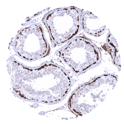
PGP9.5 / UCHL1 (MSVA-905R)
Recombinant Rabbit monoclonal / IgG 1:100 – 1:200 Research Use Only Cytoplasmic. Endoplasmic Reticulum membrane. Human MSVA-905R Gracile Axonal Dystrophy; Neuron Cytoplasmic Protein 9.5; Park5; Parkinson Disease 5; PGP95; Protein Gene Product 9.5; Ubiquitin Carboxyl-terminal Esterase L1; Ubiquitin Carboxyl- terminal Hydrolase Isozyme L1; Ubiquitin Thioesterase L1; Ubiquitin Thiolesterase L1 Pancreas: Islets cells and interspersed nerve fibres should show strong PGP9.5 staining. Pancreas: Acinar cells and excretory ducts should not show any PGP9.5 immunostaining. PGP9.5 is expressed in neuronal cells and key component of the ubiquitination/deubiquitination system. PGP9.5, also termed Ubiquitin C-terminal hydrolase (UCH)-L1 belongs to the Ub C- terminal hydrolases (UCHs) representing one of five sub-families of de-ubiquitination enzymes (DUBs). PGP9.5/UCH-L1 is thus an important component of the ubiquitination/deubiquitination system and plays a role in the posttranslational modification of proteins including protein degradation, relocation and functional alteration. PGP9.5 is one of the most prevalent proteins in the brain and makes up for 1-2% of the brain protein mass. It occurs in the cytoplasm and axons of neurons. As part of the axonal skeleton, it plays a role in axonal transport. Elevated serum PGP9.5 is a valuable marker for traumatic brain injury, and PGP9.5 serum levels correlate with the severity of injury and clinical outcome. Altered function and expression of UCH-L1 has been linked to neurodegenerative disease, cancer and other diseases. Several mutations and functional alterations of PGP9.5 have been found to be linked to neurodeg... The strongest PGP9.5 immunostaining is seen in all neuronal cells (brain and ganglia) and corresponding axons in the brain, neurohypophysis and in peripheral nerves. A similarly strong staining occurs in spermatogonia of the testis, the adrenal medulla, islet cells of the pancreas, and in corpora lutea of the ovary. A moderate to strong PGP9.5 positivity is also seen in Leydig cells, the theca cell layer of follicular cysts, ovarian stroma, the adenohypophysis, and in some scattered (neuroendocrine?) cells in the thymus. In several epithelial tissues, scattered PGP9.5 positive cells or groups of cells which often show a mosaic pattern with high variability of the expression levels between neighboring cells can occur. These include respiratory epithelium, fallopian tube, cauda epididymis, and (rarely) the prostate. A weak PGP9.5 staining occurs in a fraction of lymphocytes, predominantly located in the germinal centres. Decidua cells show a variable level of PGP9.5 expression. These fin... High level PGP9.5 expression is common in all kinds of neural and neuroendocrine tumors but can also occur at variable frequency in a large number of additional tumor types. The TCGA findings on PGP9.5 / UCHL1 RNA expression in different tumor categories have been summarized in the Human Protein Atlas. Laryngeal squamous cell carcinoma showing strong PGP9.5 positivity in tumor cells. Merkel cell carcinoma of the skin showing a strong PGP9.5 immunostaining of tumor cells. Testicular seminoma showing a strong PGP9.5 immunostaining in tumor cells. Cancer tissue gallery PGP9.5/UCH-L1 (MSVA-905R) publication summary Relevant publication: Scherzai et al. “PGP9.5 expression in human tumors: A tissue microarray study on 13,920 tumors from 120 different tumor entities “ Published in Pathol Res Pract. 2024 Oct 28;264:155676. doi: 10.1016/j.prp.2024.155676. Epub ahead of print. PMID: 39520970. A total of 10’942 tumors from 120 different tumor categories were successfully analyzed by using the following protocol: Heat-induced antigen retrieval for 5 minutes in an autoclave at 121°C in pH 7,8 Target Retrieval Solution buffer. MSVA-905R, at a dilution of 1:150 at 37°C for 60 minutes. Visualization of bound antibody by the EnVision Kit (Dako, Agilent). This protocol was also used for all stainings depicted in our tumor and normal tissue galleries. Overall, at least an occasional weak PGP9.5 positivity was detected in 109 of 120 tumor types with 87 tumor categories showing at least one strongly positive case. PGP9.5 positivity was most frequently seen in pheochro... IHC users have different preferences on how the stains should look like. Some prefer high staining intensity of the target stain and even accept some background. Others favor absolute specificity and lighter target stains. Factors that invariably lead to more intense staining include higher concentration of the antibody and visualization tools, longer incubation time, higher temperature during incubation, higher temperature and longer duration of the heat induced epitope retrieval (slide pretreatment). The impact of the pH during slide pretreatment has variable effects and depends on the antibody and the target protein. All images and data shown here and in our image galleries are obtained by the manual protocol described below. Other protocols resulting in equivalent staining are described as well. Manual protocol Freshly cut sections should be used (less than 10 days between cutting and staining). Heat-induced antigen retrieval for 5 minutes in an autoclave at 121°C in pH 7,8 Target ... The clinical/prognostic role of PGP9.5 expression needs to be evaluated in cancer. Visualization and quantification of nerve fibres and ganglions in the colon. Visualization and quantification of undifferentiated spermatogonia in the testis. There are two ways how the specificity of antibodies can be documented for immunohistochemistry on formalin fixed tissues. These are: 1. comparison with a second independent method for target expression measurement across a large number of different tissue types (orthogonal strategy), and 2. Comparison with one or several independent antibodies for the same target and showing that all positive staining results are also seen with other antibodies for the same target (independent antibody strategy). Orthogonal validation: For the antibody MSVA-905R specificity is suggested by the strong concordance of the immunostaining data with data from three independent RNA screening studies, including the Human Protein Atlas (HPA) RNA-seq tissue dataset, the FANTOM5 project, and the Genotype-Tissue Expression (GTEx) project, which are all summarized in the Human Protein Atlas (Tissue expression PGP9.5) Immunostaining by using MSVA-905R was almost exclusively detected in organs with documented PGP9.5...