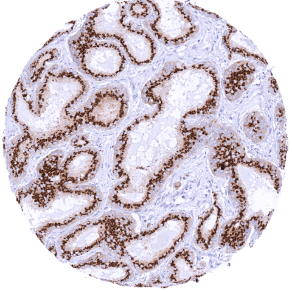
Prostein (MSVA-460R)
Recombinant Rabbit monoclonal / IgG 1:100 – 1:200 Research Use Only Membranous Human MSVA-460R SLC45A3, PCANAP6, Prostate Cancer Associated Protein 6, IPCA-6 Stomach mucosa: At least a moderate, granular, cytoplasmic prostein positivity should be seen in surface epithelial cells. Stomach mucosa: Prostein immunostaining should be absent in gastric glands. Prostein is a protein with characteristic staining pattern predominantly expressed in normal and neoplastic epithelium of the prostate. Prostein (P501S), also termed solute carrier family 45 member 3 (SLC45A3) is a 553 amino acid protein encoded by the SLC45A3 gene located at chromosome 1q32-q42. Prostein is predominantly expressed in the prostate. Its expression is androgen regulated but its function is unknown. In prostate cancer, prostein acts as the second most common 5′ partner gene in ERG rearrangements. In the brain, prostein is involved in the regulation of the lipid metabolism of oligodendrocytes and myelin. Prostein staining is always granular, cytoplasmic and predominantly perinuclear (endoplasmatic reticulum pattern). It is particularly strong in acinar cells of the prostate and occurs at lesser intensity in other cell types (shown below). Images describing the Prostein staining pattern in normal tissues obtained by the antibody MSVA-460R are shown in our “ Normal Tissue Gallery ”. Brain Cerebrum Negative. Cerebellum Negative. Endocrine Tissues Thyroid Negative. Parathyroid Negative. Adrenal gland Negative. Pituitary gland Moderate cytoplasmic prostein staining in a subset of epithelial cells of the adenohypophysis. Respiratory system Respiratory epithelium Moderate granular perinuclear cytoplasmic prostein staining in goblet cells of the respiratory epithelium. Lung Negative. Gastrointestinal Tract Salivary glands Negative. Esophagus Negative. Stomach Moderate granular perinuclear cytoplasmic prostein staining in surface epithelial cells. Duodenum Negative. Small intestine Negative. Ap... Prostein predominantly occurs in prostatic adenocarcinomas, but expression can be also seen in various other cancer types. The TCGA findings on Prostein RNA expression in different tumor categories have been summarized in the Human Protein Atlas. Prostate – Adenocarcinoma (Gleason 5+5=10) with strong Prostein immunostaining of tumor cells Stomach – Gastric adenocarcinoma showing weak granular perinuclear Prostein staining of most tumor cells Prostate – Adenocarcinoma (Gleason 3+3=6) with strong Prostein staining of tumor cells Cancer tissue gallery No data available at the moment IHC users have different preferences on how the stains should look like. Some prefer high staining intensity of the target stain and even accept some background. Others favor absolute specificity and lighter target stains. Factors that invariably lead to more intense staining include higher concentration of the antibody and visualization tools, longer incubation time, higher temperature during incubation, higher temperature and longer duration of the heat induced epitope retrieval (slide pretreatment). The impact of the pH during slide pretreatment has variable effects and depends on the antibody and the target protein. All images and data shown here and in our image galleries are obtained by the manual protocol described below. Other protocols resulting in equivalent staining are described as well. Manual protocol Freshly cut sections should be used (less than 10 days between cutting and staining). Heat-induced antigen retrieval for 5 minutes in an autoclave at 121°C in pH 7,8 Target ... The diagnostic utility of prostein for assuring a prostatic origin of cancer should be evaluated as compared to other prostate markers such as PSA and PSMA. The prognostic role of different levels of prostein expression in prostate cancer should be investigated. Given the expression of prostein in few non-prostatic tissues, its expression in corresponding tumors would be of interest. The function of prostein is unclear. The utility of prostein as a therapeutic target should be evaluated. There are two ways how the specificity of antibodies can be documented for immunohistochemistry on formalin fixed tissues. These are: 1. Comparison with a second independent method for target expression measurement across a large number of different tissue types (orthogonal strategy), and 2. Comparison with one or several independent antibodies for the same target and showing that all positive staining results are also seen with other antibodies for the same target (independent antibody strategy). Orthogonal validation: For the antibody MSVA-460R specificity is suggested by the strong concordance of the immunostaining data with data from three independent RNA screening studies, including the Human Protein Atlas (HPA) RNA-seq tissue dataset, the FANTOM5 project, and the Genotype-Tissue Expression (GTEx) project, which are all summarized in the Human Protein Atlas (Tissue expression Prostein) . The strongest immunostaining by MSVA-460R is seen in the prostate, the organ, with the highest...