PSAT1 (HMV331)
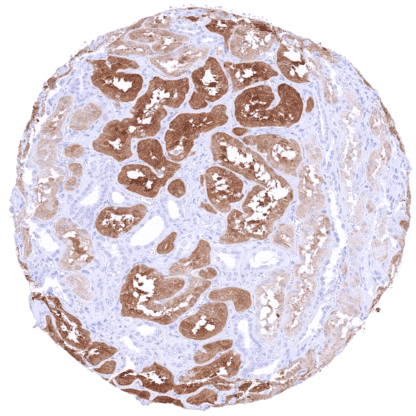
Recombinant Rabbit monoclonal / IgG 1:100 – 1:200 Research Use Only Intracellular Human HMV331 phosphoserine aminotransferase 1,EPIP,NLS2,PSA,PSAT,PSATD Kidney: A moderate to strong cytoplasmic and nuclearPSAT1staining must be seen in a fraction of tubulus cells. Lymph node: Lymphocytes should bePSAT1negative. PSAT1 is a rate limiting enzyme for glycine synthesis. Phosphoserine aminotransferase 1 (PSAT1) is coded by the PSAT1 gene at chromosome 9q21.2. It is a pivotal enzyme to produce serine and α-ketoglutarate, two critical metabolites for both carbon metabolism and the tricarboxylic acid cycle. PSAT1 is a rate limiting enzyme in the serine-glycine synthesis pathway which produces glycine, an essential nutrient for proliferating normal and neoplastic cells. Multiple studies have suggested that PSAT1 overexpression causes increased tumor cell proliferation, tumor progression, and poor patient prognosis in several different cancer types. Germ line PSAT1 mutations are among the causes for serine deficiency disorders which constitute inherited metabolic diseases with a broad phenotypic spectrum including the Neu–Laxova syndrome. In normal tissues, PSAT1 is mainly expressed in the brain, the kidney, and the pancreas but it also occurs in other cell types. Among tumors, PSAT1 expression is most often seen in brain, ovarian and endometrial carcinomas... Images describing the PSAT1 staining pattern in normal tissues obtained by the antibody HMV331 are shown in our “ Normal Tissue Gallery ”. Brain Cerebrum Strong, nuclear and cytoplasmic PSAT1 staining of astrocytes. Cerebellum Nuclear and cytoplasmic PSAT1 staining is particularly strong in a subset of glial cells between the molecular and the granule cell layer. Endocrine Tissues Thyroid Negative. Parathyroid Weak to moderate, predominantly cytoplasmic PSAT1 staining of a small subset of cells (not in all samples). Adrenal gland Weak to moderate, predominantly cytoplasmic PSAT1 staining of a subset of cells. Pituitary gland Strong, predominantly cytoplasmic PSAT1 staining of a small subset of epithelial cells in the anterior lobe. Respiratory system Respiratory epithelium Negative. Lung Negative. Gastrointestinal Tract Salivary glands Cytoplasmic and nuclear PSAT1 staining of variable intensity can occur in serous glandular cells Esophagus Moderate, nuclear and cytoplasmic PSAT1 stain... PSAT1 is most often expressed in brain, ovarian and endometrium carcinomas, but PSAT1 expression can also occur in tumors of various other organs. The TCGA findings on PSAT1 RNA expression in different tumor categories have been summarized in the Human Protein Atlas. Lung – Squamous cell carcinoma with moderate to strong, predominantly nuclear PSAT1 staining of tumor cells Thyroid – PSAT1 negative papillary carcinoma Prostate – Adenocarcinoma (Gleason 5+5=10) with strong PSAT1 staining of tumor cells Cancer tissue gallery No data available at the moment IHC users have different preferences on how the stains should look like. Some prefer high staining intensity of the target stain and even accept some background. Others favor absolute specificity and lighter target stains. Factors that invariably lead to more intense staining include higher concentration of the antibody and visualization tools, longer incubation time, higher temperature during incubation, higher temperature and longer duration of the heat induced epitope retrieval (slide pretreatment). The impact of the pH during slide pretreatment has variable effects and depends on the antibody and the target protein. All images and data shown here and in our image galleries are obtained by the manual protocol described below. Other protocols resulting in equivalent staining are described as well. Manual protocol Freshly cut sections should be used (less than 10 days between cutting and staining). Heat-induced antigen retrieval for 5 minutes in an autoclave at 121°C in pH 7,8 Target ... The diagnostic and prognostic relevance of PSAT1 IHC in tumors and in preneoplastic disease is unresolved. The function of PSAT1 in cancer cells is unknown. PSAT1 may represent a therapeutic target, at least in tumors with p53-72Pro variant. There are two ways how the specificity of antibodies can be documented for immunohistochemistry on formalin fixed tissues. These are: 1. Comparison with a second independent method for target expression measurement across a large number of different tissue types (orthogonal strategy), and 2. Comparison with one or several independent antibodies for the same target and showing that all positive staining results are also seen with other antibodies for the same target (independent antibody strategy). Orthogonal validation: For the antibody HMV331, specificity is supported by the strong concordance with RNA expression data derived from three independent RNA screening studies, including the Human Protein Atlas (HPA) RNA-seq tissue dataset, the FANTOM5 project, and the Genotype-Tissue Expression (GTEx) project, which are all summarized in the Human Protein Atlas (Tissue expression PSAT1) . A PSAT1 staining was observed in all organs with a significant PSAT1 RNA expression (Brain, esophagus, ...