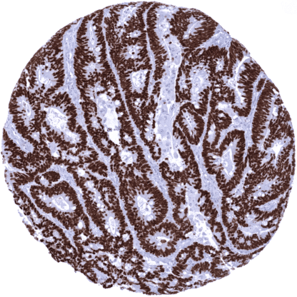
SATB2 (MSVA-702R)
Recombinant Rabbit monoclonal / IgG 1:100 – 1:200 Research Use Only Nuclear Human MSVA-702R DNA-binding protein SATB2; GLSS; SATB homeobox 2; Special AT-rich sequence-binding protein 2 Colon: A strong nuclear staining should be seen in virtually all columnar epithelial cells. Colon: SATB2 staining should be absent in stromal and smooth muscle cells. SATB2 is expressed in colorectal epithelial cells, osteoclasts, and the brain. Special AT-rich sequence-binding protein 2 (SATB2) is a nuclear protein with a molecular weight of 82.5 kDa encoded by the SATB2 gene at chromosome 2q33. SATB2 specifically binds to so-called matrix attachment regions (MARs) of the DNA, where it induces local chromatin-loop remodeling that impacts the transcriptional activity of the affected genes. Attached SATB2 recruits additional chromatin remodeling enzymes including histone deacetylases (HDACs) and histone acetyltransferases (HATs). Genes regulated by SATB2 include programs required for neuronal, skeletal, and osteoblast development. SATB2 staining pattern in Normal Tissues with antibody MSVA-702R (images are shown in our “Normal Tissue Gallery”) Brain Cerebrum Weak to moderate nuclear SATB2 staining of neurons. An additional fibrillar staining may represent a (tolerable) cross-reactivity. Cerebellum Weak to moderate nuclear SATB2 staining of neurons. Endocrine Tissues Thyroid Negative. Parathyroid Negative. Adrenal gland Negative. Pituitary gland Negative. Respiratory system Respiratory epithelium Negative. Lung Negative. Gastrointestinal Tract Salivary glands Negative. Esophagus Negative. Stomach Negative. Duodenum Negative. Small intestine Weak to moderate nuclear SATB2 staining of all epithelial cells. Appendix Strong nuclear SATB2 staining of all epithelial cells. Colon Strong nuclear SATB2 staining of all epithelial cells. Rectum Strong nuclear SATB2 staining of all epithelial cells. Liver Negative. Gallbladder Negative. Pancreas Negative. Genitourinary Kidney Weak to moderate nuclear SATB2 staining of a frac... A positive SATB2 immunostaining is most commonly seen in adenocarcinomas and neuroendocrine tumors derived from the colorectum or appendix, osteosarcomas, and in Merkel cell cancer. A positive SATB2 immunostaining, often at lower intensity, has also been described to occur in various other tumor entities. The TCGA findings on SATB2 RNA expression in different tumor categories have been summarized in the Human Protein Atlas. Colorectal adenocarcinoma showing strong SATB2 immunostaining of all tumor cells. Esophageal squamous cell carcinoma depicting a strong SATB2 positivity of the majority of tumor cells. Endometroid endometrium carcinoma with weak to moderate SATB2 immunostaining of the majority of tumor cells. Cancer tissue gallery SATB2 (MSVA-702R) publication summary Relevant publication: Dum et al. “ SATB2 Expression in Human Tumors: A Tissue Microarray Study on More Than 15 000 Tumors.” A total of 11’678 tumors were successfully analyzed from 120 different tumor categories by using the following protocol: Heat-induced antigen retrieval for 5 minutes in an autoclave at 121°C in pH7,8 Target Retrieval Solution buffer. MSVA-702R at a dilution of 1:100 at 37°C for 60 minutes. Visualization of bound antibody by the EnVision Kit (Dako, Agilent). This protocol was also used for all stainings depicted in our tumor and normal tissue galleries. In this study, at least one positive case was seen in 89 (74%) of 120 tumor categories and 38 (32%) tumor categories included at least one case with strong positivity. The highest rates of positive staining and the highest levels of expression were found in adenomas and adenocarcinomas of the colorectum, various subtypes of neuroendocrine tumors of the colorectum and the appendi... IHC users have different preferences on how the stains should look like. Some prefer high staining intensity of the target stain and even accept some background. Others favor absolute specificity and lighter target stains. Factors that invariably lead to more intense staining include higher concentration of the antibody and visualization tools, longer incubation time, higher temperature during incubation, higher temperature and longer duration of the heat induced epitope retrieval (slide pretreatment). The impact of the pH during slide pretreatment has variable effects and depends on the antibody and the target protein. All images and data shown here and in our image galleries are obtained by the manual protocol described below. Other protocols resulting in equivalent staining are described as well. -Manual protocol Freshly cut sections should be used (less than 10 days between cutting and staining). Heat-induced antigen retrieval for 5 minutes in an autoclave at 121°C in pH 7,8 Target... The diagnostic utility of SATB2 expression analysis should be further investigated in a large cohort of tumors from different entities The prognostic role of SATB2 expression in gastrointestinal adenocarcinomas is unclear. There are two ways how the specificity of antibodies can be documented for immunohistochemistry on formalin fixed tissues. These are: 1. Comparison with a second independent method for target expression measurement across a large number of different tissue types (orthogonal strategy), and 2. Comparison with one or several independent antibodies for the same target and showing that all positive staining results are also seen with other antibodies for the same target (independent antibody strategy). Orthogonal validation: For the antibody MSVA-702R specificity is suggested by the strong concordance of its staining results with RNA expression data derived from three independent RNA screening studies, including the Human Protein Atlas (HPA) RNA-seq tissue dataset, the FANTOM5 project, and the Genotype-Tissue Expression (GTEx) project, which are all summarized in the Human Protein Atlas (Tissue expression SATB2) . Immunostaining by using MSVA-702R is only detected in organs with documented ...