SMARCA2 (HMV337)
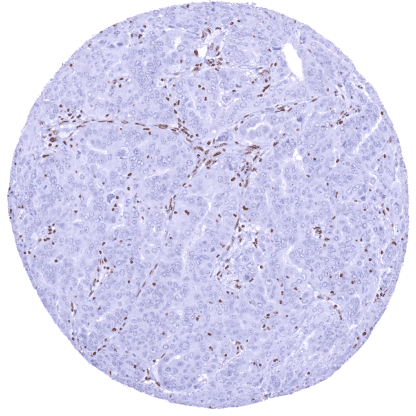
Recombinant Rabbit monoclonal / IgG 1:100 – 1:200 Research Use Only Nucleus Human HMV337 SWI/SNF related, matrix associated, actin dependent regulator of chromatin, subfamily a, member 2 , BAF190 , BRM , NCBRS , SNF2 , SNF2L2 , SNF2LA , SWI2 , Sth1p , hBRM , hSNF2a Colon: All cells should display a moderate to strong SMARCA2 immunostaining although the nuclear staining intensity decreases somewhat from the crypt base to the surface epithelium. Testis: A subset of intratubular cells (Sertoli cells) must be SMARCA2 negative. SMARCA2 is a key protein of the SWI/SNF complex. SMARCA2 (SWI/SNF ‐ related matrix ‐ associated actin ‐ dependent regulator of chromatin subfamily A member 2) is coded by the SMARCA2 gene located at chromosome 9p24.3. Along with SMARCA4, SMARCA2 is one of two mutually exclusive, exchangeable DNA-dependent ATPases, which constitute the enzymatic motor of a polymorphic family of SWI/SNF complexes which also include 8–15 further subunits (). SWI/SNF can modify the transcription of genes by altering the chromatin structure surrounding them. SMARCA2 is ubiquitously expressed in the nuclei of normal cells. Mutations in individual members of the SWI/SNF family together represent one of the most common genetic alterations in cancer, observed in about 20% of cases. According to public databases, mutations of SMARCA2 are rare but SMARCA2 downregulation in cancer can occur due to alternative mechanisms. Targeting SMARCA2 may cause synthetic lethality in SMARCA4 deficient cancers. A nuclear SMARCA2 immunostaining is seen in all normal tissues, and in most cell types. The intensity of nuclear staining is somewhat variable between tissues. Images describing the SMARCA2 staining pattern in normal tissues obtained by the antibody HMV337 are shown in our “ Normal Tissue Gallery ”. Brain Cerebrum Strong nuclear SMARCA2 staining of most glia cells while staining is faint or absent in neurons. Cerebellum Strong nuclear SMARCA2 staining of most glia cells while staining is lacking in granule cells and in Purkinje cells. Endocrine Tissues Thyroid Nuclear SMARCA2 staining of all cells. Parathyroid Nuclear SMARCA2 staining of all cells. Adrenal gland Nuclear SMARCA2 staining of all cells. Pituitary gland In the adenohypophysis the nuclear SMARCA2 staining varies between individual epithelial cells ranging from strongly positive to negative. Most pituicytes show a strong nuclear SMARCA2 staining in the neurohypophysis. Respiratory system Respiratory epithelium Nuclear SMARC... Nuclear SMARCA2 expression – at various levels – is seen in the vast majority of cancers. A complete loss of SMARCA2 staining can also occur. It is seen in a small fraction of cases in many different tumor entities. The TCGA findings on SMARCA2 RNA expression in different tumor categories have been summarized in the Human Protein Atlas. SMARCA2 deficient adenocarcinoma of
the papilla Vaterii. Colorectal adenocarcinoma with strong
SMARCA2 staining of tumor cells. Pulmonary adenocarcinoma with a complete
SMARCA2 loss. Cancer tissue gallery No data available at the moment IHC users have different preferences on how the stains should look like. Some prefer high staining intensity of the target stain and even accept some background. Others favor absolute specificity and lighter target stains. Factors that invariably lead to more intense staining include higher concentration of the antibody and visualization tools, longer incubation time, higher temperature during incubation, higher temperature and longer duration of the heat induced epitope retrieval (slide pretreatment). The impact of the pH during slide pretreatment has variable effects and depends on the antibody and the target protein. All images and data shown here and in our image galleries are obtained by the manual protocol described below. Other protocols resulting in equivalent staining are described as well. Manual protocol Freshly cut sections should be used (less than 10 days between cutting and staining). Heat-induced antigen retrieval for 5 minutes in an autoclave at 121°C in pH 7,8 Target ... The SWI/SNF complex has great importance in cancer biology. Inactivation of one of 29 genes coding for the components of the complex due to a mutation occurs in >20% of all cancers. The functions and interactions of the individual components such as SMARCA2 are not yet fully understood. The expression levels of SMARCA2 (absent, reduced, normal, increased) may have a biological/clinical relevance in cancer. The ratio of SMARCA2/SMARCA4 expression may be clinically important in cancer. There are two ways how the specificity of antibodies can be documented for immunohistochemistry on formalin fixed tissues. These are: 1. Comparison with a second independent method for target expression measurement across a large number of different tissue types (orthogonal strategy), and 2. Comparison with one or several independent antibodies for the same target and showing that all positive staining results are also seen with other antibodies for the same target (independent antibody strategy). Orthogonal validation: Because of the ubiquitous expression of SMARCA2, orthogonal validation is not applicable. Comparison of antibodies: True expression of SMARCA2 in cells showing SMARCA2 immunostaining by HMV337 is suggested by the staining distribution (almost all cells are positive) and the confirmation of tissue specific variations of the staining intensity by a comparison with a commercially available independent second antibody (termed “validation antibody”). This validation staining...