SMARCA4 / BRG1 (MSVA-397R)
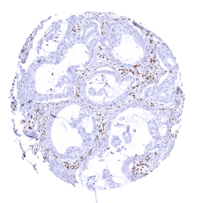
Recombinant Rabbit monoclonal / IgG 1:100 – 1:200 Research Use Only Nucleus Human MSVA-397R ATP dependent helicase SMARCA4; BAF190A; Brahma protein like 1; BRM/SWI2 related gene 1; global transcription activator snf2l4; Homeotic gene regulator; hSNF2b; Mitotic growth and transcription activator; MRD16; Nuclear protein GRB1; Protein brahma homolog 1; RTPS2; SMARCA4;; SNF2LB; SWI2; Transcription activator BRG1 Kidney: All cells should display an at least moderate nuclear SMARCA4 immunostaining. A tumor (or cell line) with documented SMARCA4 expression loss should be used as a “negative” control (as SMARCA4 is expressed in all normal tissues) SMARCA4 is a key protein of the SWI/SNF complex. SMARCA4 (SWI/SNF ‐ related matrix ‐ associated actin ‐ dependent regulator of chromatin subfamily A member 4) also termed BRG1 (Brahma-related gene-1) is coded by the SMARCA4 gene located at chromosome 19p13.2. SMARCA4 is one of two mutually exclusive DNA-dependent ATPases, along with SMARCA2, involved in transcriptional regulation of gene expression. SMARCA4 exerts its regulatory role as a part of multiple major components of the large ATP-dependent chromatin remodeling complex SWI/SNF, which can modify the transcription of genes by altering the chromatin structure surrounding them. SMARCA4 is ubiquitously expressed in the nuclei of all normal cells. Mutations in individual members of the SWI/SNF family together represent one of the most common genetic alterations in cancer, observed in about 20% of cases. Among these, SMARCA4 is the most frequently mutated family member. SMARCA4 mutations are thought to occur in 5-7% of all malignant tumors. SMARCA4 is most commonly affected in lung ... A nuclear SMARCA4 immunostaining is seen in all normal tissues, sometimes accompanied by some cytoplasmic staining. The intensity of nuclear staining is somewhat variable between tissues. Images describing the SMARCA4 staining pattern in normal tissues obtained by the antibody MSVA-397 R are shown in our “ Normal Tissue Gallery ”. Brain Cerebrum Nuclear SMARCA4 staining of variable intensity. Cerebellum Nuclear SMARCA4 staining of variable intensity. Endocrine Tissues Thyroid Nuclear SMARCA4 staining. Parathyroid Nuclear SMARCA4 staining. Adrenal gland Nuclear SMARCA4 staining. Pituitary gland Nuclear SMARCA4 staining of all cells of adenohypophysis and neurohypophysis. Respiratory system Respiratory epithelium Nuclear SMARCA4 staining. Lung Nuclear SMARCA4 staining. Gastrointestinal Tract Salivary glands Nuclear SMARCA4 staining. Esophagus Nuclear SMARCA4 staining. Stomach Nuclear SMARCA4 staining. Duodenum Nuclear SMARCA4 staining. Small intestine Nuclear SMARCA4 staining. Appendix ... SMARCA4 mutation and consecutive loss of expression is implicated in the pathogenesis of several malignancies. Loss of SMARCA4 mutation is particularly common in small cell carcinoma of the ovary, hypercalcemic type (SCCOHT) and in lung cancer. SMARCA4 expression loss can also be found in various other malignant tumors. Note: Most SMARCA4 negative lung carcinomas are “regular” adenocarcinomas. The diagnosis of “ SMARCA4-deficient undifferentiated tumor (SMARCA4-dUT) of the lung” is reserved for cancers lacking unequivocal glandular differentiation. The TCGA findings on SMARCA4 RNA expression in different tumor categories have been summarized in the Human Protein Atlas. Mucoepidermoid carcinoma with complete loss of SMARCA4 staining in all tumor cells. Staining is retained in all non-neoplastic cells Adenocarcinoma with strong SMARCA4 immunostaining of all tumor cells Hepatocellular carcinoma with distinct nuclear SMARCA4 positivity of tumor cells Cancer tissue gallery No data available at the moment IHC users have different preferences on how the stains should look like. Some prefer high staining intensity of the target stain and even accept some background. Others favor absolute specificity and lighter target stains. Factors that invariably lead to more intense staining include higher concentration of the antibody and visualization tools, longer incubation time, higher temperature during incubation, higher temperature and longer duration of the heat induced epitope retrieval (slide pretreatment). The impact of the pH during slide pretreatment has variable effects and depends on the antibody and the target protein. All images and data shown here and in our image galleries are obtained by the manual protocol described below. Other protocols resulting in equivalent staining are described as well. Manual protocol Freshly cut sections should be used (less than 10 days between cutting and staining). Heat-induced antigen retrieval for 5 minutes in an autoclave at 121°C in pH 7,8 Target ... The SWI/SNF complex has great importance in cancer biology. Inactivation of one of 29 genes coding for the components of the complex due to a mutation occurs in >20% of all cancers. The functions of the individual components such as SMARCA4 are not yet fully understood. Alterations of SWI/SNF complex and reduced expression of SMARCA4 may result in increased likelihood for a favorable response to Immune checkpoint Inhibitors. The clinical and biological significance of altered SMARCA4 expression patterns (mosaic pattern!) is not understood. The expression levels of SMARCA4 (reduced, normal, increased) may have a biological/clinical relevance in cancer. There are two ways how the specificity of antibodies can be documented for immunohistochemistry on formalin fixed tissues. These are: 1. Comparison with a second independent method for target expression measurement across a large number of different tissue types (orthogonal strategy), and 2. Comparison with one or several independent antibodies for the same target and showing that all positive staining results are also seen with other antibodies for the same target (independent antibody strategy). Orthogonal validation: Because of the ubiquitous expression of SMARCA4, orthogonal validation is not applicable. Comparison of antibodies: True expression of SMARCA4 in cells showing SMARCA4 immunostaining is suggested by the staining distribution (all cells are positive) and a comparison with a commercially available independent second antibody (termed “validation antibody” ) showing identical staining patterns including a loss in tumors with SMARCA4 expression loss obtained by MSVA-397R. An...