Thyroglobulin (MSVA-189R)
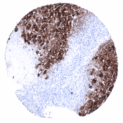
Recombinant Rabbit monoclonal / IgG 1:100 – 1:200 Research Use Only Cytoplasmic Human MSVA-189R AITD3, hTG, TDH3, Tg, Tgn Thyroid: A strong staining is seen in follicular cells and the colloid. The stroma is typically also stained due to a contamination artifact. Colon: All cells must not show any staining. Thyroglobulin is a 660 kDa glycoprotein encoded by the TG gene located at human chromosome 8q24.22. The glycoprotein is predominantly expressed by follicular cells of the thyroid gland and assembles into homodimers which are secreted into the follicular lumen. Thyroglobulin is the substrate for the synthesis of thyroxine and triiodothyronine, two major hormones of the thyroid gland, and serves as a storage protein for the inactive forms of these hormones in the follicular colloid. Polymorphisms of the TG gene have been linked to susceptibility of autoimmune thyroid diseases including Hashimoto thyroiditis.[1] Thyroglobulin is a 660 kDa glycoprotein encoded by the TG gene located at human chromosome 8q24.22. The glycoprotein is predominantly expressed by follicular cells of the thyroid gland and assembles into homodimers which are secreted into the follicular lumen. Thyroglobulin is the substrate for the synthesis of thyroxine and triiodothyronine, two major hormones of the thyroid gland, and serves as a storage protein for the inactive forms of these hormones in the follicular colloid. Polymorphisms of the TG gene have been linked to susceptibility of autoimmune thyroid diseases including Hashimoto thyroiditis. [1] [1] Steurer et al.: “Immunohistochemically detectable thyroglobulin expression in extrathyroidal cancer is 100% specific for thyroidal tumor origin” . Published in Annals of Diagnostic Pathology, Volume 54, October 2021, 151793 In normal tissues, thyroglobulin immunostaining is exclusively seen in thyroid tissue. In the thyroid, immunostaining often not only involves follicular epithelial cells but also stroma and other cell types. This is due to the abundance of thyreoglobulin in the thyroid. Thyroglobulin makes up for about half of the protein mass of the thyroid. In case of immunostaining, some thyroglobulin spill-over occurs resulting in a staining of follicle-adjacent tissues. These findings are largely comparable to the data described in the Human Protein Atlas (Tissue expression Thyroglobulin) . Suggested positive tissue control : Thyroid: A strong staining is seen in follicular cells and the colloid. The stroma is typically also stained due to a contamination artifact. Suggested negative tissue control: Colon: All cells must not show any staining. Thyroid gland – Very intense thyroglobulin immunostaining in follicular cells. Due to the large quantity of thyroglobulin in a normal thyroid (50% of the p... Thyroglobulin expression is only found in tumors derived from the thyroid and in metastases from extrathyroidal tumors to the thyroid. In the case of metastases to the thyroid, tissue contamination by colloid from potentially destructed adjacent follicles is likely to cause unexpected positivity in a fraction of cases. For the same reason thyroglobulin immunostaining can also be seen in medullary (or anaplastic) carcinomas of the thyroid, which do not express thyroglobulin. Detailed data on Thyroglobulin staining by MSVA-189R obtained from an analysis of >9000 tumors from 109 different tumor types and subtypes have recently been published Steurer et al.: “Immunohistochemically detectable thyroglobulin expression in extrathyroidal cancer is 100% specific for thyroidal tumor origin” . – Detailed data below “Compatibility of Antibodies” The TCGA findings on Thyroglobulin RNA expression in different tumor categories have been summarized in the Human Protein Atlas. Follicular thyroid cancer... Thyroglobulin (MSVA-189R) publication summary Paper used for data compilation: Steurer et al.: “Immunohistochemically detectable thyroglobulin expression in extrathyroidal cancer is 100% specific for thyroidal tumor origin” . Published in Annals of Diagnostic Pathology, Volume 54, October 2021, 151793 In this study, a total of 9974 tumors were analyzed from 109 different tumor categories by using the following protocol: Heat-induced antigen retrieval for 5 minutes in an autoclave at 121°C in pH 6 Target Retrieveal Solution buffer. MSVA-189R at a dilution of 1:135 at 37°C for 60 minutes. Visualization of bound antibody by the EnVision Kit (Dako, Agilent). This protocol was also used for all stainings depicted in our tumor and normal tissue galleries. The distribution of positive staining results is shown in an “organ-systematic” and in a “ranking order” figure below (images based on a compilation of data from Steurer et al. ). A positive thyroglobulin immunostaining was only seen in tum... IHC users have different preferences on how the stains should look like. Some prefer high staining intensity of the target stain and even accept some background. Others favor absolute specificity and lighter target stains. Factors that invariably lead to more intense staining include higher concentration of the antibody and visualization tools, longer incubation time, higher temperature during incubation, higher temperature and longer duration of the heat induced epitope retrieval (slide pretreatment). The impact of the pH during slide pretreatment has variable effects and depends on the antibody and the target protein. All images and data shown here and in our image galleries are obtained by the manual protocol described below. Other protocols resulting in equivalent staining are described as well. Manual protocol Freshly cut TMA sections were deparaffinized and exposed to heat-induced antigen retrieval for 5 minutes in an autoclave at 121°C in pH 6 Target Retrieval Solution buffer. A... No data available at the moment. Specificity of MSVA-189R is documented by strong positive staining thyroid follicular cells in combination with complete absence of any thyroglobulin immunostaining in any other normal tissues. Normal tissue gallery