TIGIT (HMV323)
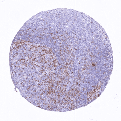
Recombinant Rabbit monoclonal / IgG 1:100 – 1:200 Research Use Only Membraneous Human HMV323 T cell immunoreceptor with Ig and ITIM domains , VSIG9 , VSTM3 , WUCAM Tonsil: A moderate to strongTIGITstaining should be seen in a subset of germinal centre cells (CD4+follicular T helper cells located in the germinal centre periphery orientated towards the tonsil surface epithelium). Subsets of other lymphocytes should also be positive but less intense. Tonsil:TIGITstaining should be absent in all epithelial cells and in most lymphocytes. TIGIT is a pivotal target in immune-oncology. TIGIT (T cell immunoreceptor with Ig and ITIM domains) is a transmembrane glycoprotein of the poliovirus receptor (PVR) family which is coded by the TIGIT gene at 3q13.31. TIGIT acts as an inhibitory immune receptor (immune checkpoint) which can bind to CD155 with high affinity, and to CD112 with lower affinity. TIGIT expression is restricted to some CD8 + cytotoxic T cells, CD4 + T helper cells, FOXP3 + regulatory T cells, and NK cells . The highest level of expression occurs in CD4 + follicular T helper cells located in the germinal centre periphery orientated towards the tonsil surface epithelium. TIGIT expression has a limiting effect on antitumoral immune reactions. TIGIT inhibition, by either genetic ablation or blocking antibodies, increases T-cell activation and proliferation in response to stimulation and consequently results in reduced tumor growth in experimental models. Various compounds targeting TIGIT have been developed (i.e. tiragolumab, domvanalimab, vibostolimab, etig... Images describing the TIGIT staining pattern in normal tissues obtained by the antibody HMV323 are shown in our “ Normal Tissue Gallery ”. Brain Cerebrum Negative. Cerebellum Negative. Endocrine Tissues Thyroid Negative. Parathyroid Negative. Adrenal gland Negative. Pituitary gland Negative. Respiratory system Respiratory epithelium Negative. Lung Negative. Gastrointestinal Tract Salivary glands Negative. Esophagus Negative. Stomach Negative. Duodenum Negative. Small intestine Negative. Appendix Negative. Colon Negative. Rectum Negative. Liver Negative. Gallbladder Negative. Pancreas Negative. Genitourinary Kidney Negative. Urothelium Negative. Male genital Prostate Negative. Seminal vesicles Negative. Testis Negative. Epididymis Negative. Female genital Breast Negative. Uterus, myometrium Negative. Uterus, ectocervix Negative. Uterus endocervix Negative. Uterus, endometrium Negative. Fallopian Tube Negative. Ovary Negative. Placenta early Negative. Placenta mature Negative. Amnion Neg... TIGIT is expressed in a variable subset of tumor associated T-cells. TIGIT expression may also occur in some T-cell lymphomas. The TCGA findings on TIGIT RNA expression in different tumor categories have been summarized in the Human Protein Atlas. Hodgkin’s lymphoma with TIGIT
positivity of a subset of tumor
infiltrating non-neoplastic
lymphocytes. Follicular B-cell lymphoma with strong
TIGIT positivity of a portion of tumor
infiltrating non-neoplastic lymphocytes. Endometrioid endometrium carcinoma
with a moderate to strong TIGIT staining
of some tumor infiltrating lymphocytes. Cancer tissue gallery No data available at the moment IHC users have different preferences on how the stains should look like. Some prefer high staining intensity of the target stain and even accept some background. Others favor absolute specificity and lighter target stains. Factors that invariably lead to more intense staining include higher concentration of the antibody and visualization tools, longer incubation time, higher temperature during incubation, higher temperature and longer duration of the heat induced epitope retrieval (slide pretreatment). The impact of the pH during slide pretreatment has variable effects and depends on the antibody and the target protein. All images and data shown here and in our image galleries are obtained by the manual protocol described below. Other protocols resulting in equivalent staining are described as well. Manual protocol Freshly cut sections should be used (less than 10 days between cutting and staining). Heat-induced antigen retrieval for 5 minutes in an autoclave at 121°C in pH 7,8 Target ... The role of TIGIT as a drug target is under scrutiny. The predictive role of TIGIT analysis in tissues is unknown. The downstream signaling of TIGIT is not fully understood. There are two ways how the specificity of antibodies can be documented for immunohistochemistry on formalin fixed tissues. These are: 1. Comparison with a second independent method for target expression measurement across many different tissue types (orthogonal strategy), and 2. Comparison with one or several independent antibodies for the same target and showing that all positive staining results are also seen with other antibodies for the same target (independent antibody strategy). Orthogonal validation: Comparison with RNA expression data is not well suited to validate immunohistochemical stainings of cell types such as hematolymphoid cells which occur in virtually all organs. Nevertheless, in agreement with data from three independent RNA screening studies, including the Human Protein Atlas (HPA) RNA-seq tissue dataset, the FANTOM5 project, and the Genotype-Tissue Expression (GTEx) project, which are all summarized in the Human Protein Atlas (Tissue expression TIGIT) , TIGIT immun...