TIM-3 (MSVA-366R)
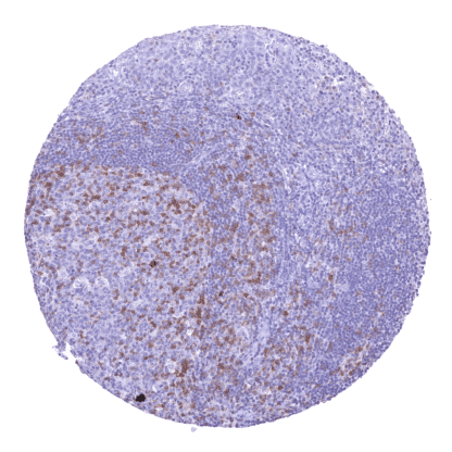
Recombinant Rabbit monoclonal / IgG 1:100 – 1:200 Research Use Only Cell Surface Human MSVA-366R CD366; HAVR2; Hepatitis A virus cellular receptor 2 (HAVCR2); Kidney injury molecule 3 (KIM3); T-cell immunoglobulin and mucin domain-containing protein 3; T-cell immunoglobulin mucin receptor 3; T-cell membrane protein 3; TIM3; TIMD3 Tonsil: A moderate to strong TIM-3 immunostaining should be seen in a significant fraction of T-cells, while only few TIM-3 positive cells are seen in the germinal centre and the mantle zone. Tonsil: Epithelial cells should be negative. TIM-3 is an immune checkpoint that constitutes a “hot topic” in immune-oncology research. TIM-3 (T-cell immunoglobulin and mucin-domain containing-3) also termed Hepatitis A virus cellular receptor 2 (HAVCR2) is a 33 kDa cell surface receptor protein coded by the TIM-3 gene on 5q33.3 . TIM-3 belongs to TIM family cell surface receptor proteins. These proteins share a similar structure with an extracellular region containing a variable immunoglobulin domain (IgV) and a glycosylated mucin domain of variable length. TIM-3 is an immune checkpoint. It mediates CD8+ T-cell exhaustion together with other inhibitory receptors such as PD-1 and LAG3. TIM-3 is primarily activated by galectin-9. The interaction with galectin-9 leads to stimulation of an influx of calcium to the intracellular space and induction of apoptosis. Other known TIM-3 ligands include phosphatidylserine (PtdSer), High Mobility Group Protein 1 (HMGB1) and Carcinoembryonic Antigen Related Cell Adhesion Molecule 1 (CEACAM1). TIM-3 is viewed as a promising drug target. Multiple phase 1/2 clinical trials with anti-... TIM-3 staining pattern in Normal Tissues with antibody MSVA-366R (images are shown in our “Normal Tissue Gallery”) Brain Cerebrum Negative. Cerebellum Negative. Endocrine Tissues Thyroid Negative. Parathyroid Negative. Adrenal gland TIM-3 positivity of some tissue associated macrophages. Pituitary gland Negative. Respiratory system Respiratory epithelium TIM-3 positivity of some tissue associated macrophages and/or lymphocytes. Lung Strong TIM-3 positivity of alveolar macrophages. Gastrointestinal Tract Salivary glands Epithelial cells are TIM-3 negative. Esophagus Epithelial cells are TIM-3 negative. Stomach Epithelial cells are TIM-3 negative. Duodenum Moderate TIM-3 staining of apical membranes of surface epithelial may reflect a (tolerable) cross-reactivity. Small intestine Moderate TIM-3 staining of apical membranes of surface epithelial may reflect a (tolerable) cross-reactivity. Appendix TIM-3 positivity of a fraction of tissue associated macrophages. Colon TIM-3 positivity of a... TIM-3 positive lymphocytes and macrophages occur at variable density in virtually all cancers. TIM-3 can also be expressed on cancer cells such as for example in a fraction of renal cell carcinomas. The TCGA findings on TIM-3 RNA expression in different tumor categories have been summarized in the Human Protein Atlas. Oral cavity- TIM3 negative squamous cell carcinoma containing numerous TIM3 positive macrophages and also lymphocytes. Thorax- TIM3 negative epitheloid mesothelioma containing numerous TIM3 positive inflammatory cells. TIM3 negative seminoma containing numerous TIM3 positive macrophages and lymphocytes. Cancer tissue gallery No data available at the moment IHC users have different preferences on how the stains should look like. Some prefer high staining intensity of the target stain and even accept some background. Others favor absolute specificity and lighter target stains. Factors that invariably lead to more intense staining include higher concentration of the antibody and visualization tools, longer incubation time, higher temperature during incubation, higher temperature and longer duration of the heat induced epitope retrieval (slide pretreatment). The impact of the pH during slide pretreatment has variable effects and depends on the antibody and the target protein. All images and data shown here and in our image galleries are obtained by the manual protocol described below. Other protocols resulting in equivalent staining are described as well. Manual protocol Freshly cut sections should be used (less than 10 days between cutting and staining). Heat-induced antigen retrieval for 5 minutes in an autoclave at 121°C in pH 7,8 Target ... TIM-3 is an important immune checkpoint, the function of which deserves further evaluation. As TIM-3 can be expressed in epithelial cells (kidney, epididymis), the expression of TIM-3 in tumor cells of cancerous tissues needs to be investigated. The clinical significance of the density of TIM-3 positive lymphocytes and macrophages needs to be investigated. There are two ways, how the specificity of antibodies can be documented for immunohistochemistry on formalin fixed tissues. These are: 1. comparison with a second independent method for target expression measurement across a large number of different tissue types (orthogonal strategy), and 2. Comparison with one or several independent antibodies for the same target and showing that all positive staining results are also seen with other antibodies for the same target (independent antibody strategy). As a standard validation process for MSVA antibodies, RNA data summarized in the protein atlas are used for comparison. However, this has limited validity for proteins that are expressed in significant subsets of lymphocytes and macrophages because these cells occur in virtually all organs. The validation of MSVA-366R is thus based on a comparison with other antibodies. The MSVA-366R findings of a significant TIM-3 protein expression in: Kupffer cells of the liver epididymis spleen vessels a...