TLE1 (MSVA-856R)
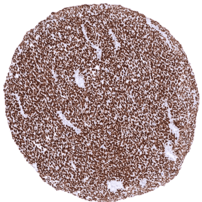
Recombinant Rabbit monoclonal / IgG 1:100 – 1:200 Research Use Only Nucleus Human MSVA-856R Enhancer of split groucho 1 (ESG1); Transducin like enhancer of split 1; Transducin-like enhancer protein 1; Enhancer of split groucho-like protein 1; GRG1; TLE1 Tonsil: A weak to moderate TLE1 staining should be seen in suprabasal cells of squamous epithelium and in a small subset of lymphocytes in germinal centres. Tonsil: The majority of lymphocytes in germinal centres should be TLE1 negative. TLE1 is Overexpressed in 80% of synovial sarcomas. Transducin-like enhancer of split 1 ( TLE1 ) is a member of the Groucho (Gro)/TLE family of transcriptional co-repressors. The TLE1 gene at chromosome 9q21.32 consists of 19 exons and encodes a 770 amino acid protein. TLE1 is a co-repressor of multiple other transcription factors. TLE1 does not bind to DNA but forms homo-oligomers and hetero-oligomers with other transcription factor proteins to inhibit their transcriptional activity. TLE1 interacts with several signal transduction pathways including the NF-kB Notch, Wnt/b-catenin, and Dpp/BMP/TGF-β signaling pathways. Accordingly, TLE1 regulates the transcriptional activity of a wide range of genes and has been implicated in embryogenesis, hematopoiesis, sex determination, neuronal and terminal epithelial differentiation, or cell proliferation and migration. TLE1 plays a role in the regulation of inflammation and in cancer progression. Images describing the TLE1staining pattern in normal tissues obtained by the antibody MSVA-856R are shown in our “ Normal Tissue Gallery ”. Brain Cerebrum Weak to moderate TLE1 staining of subsets of glial and neuronal cells. Cerebellum Weak to moderate TLE1 staining of subsets of glial and neuronal cells. Endocrine Tissues Thyroid Weak TLE1 staining of follicular and endothelial cells. Parathyroid TLE1 staining is largely limited to endothelial cells. Adrenal gland Weak TLE1 staining of a fraction of adrenocortical and medullary cells. Pituitary gland Weak to moderate TLE1 staining of a fraction of epithelial cells of the adenohypophysis. Respiratory system Respiratory epithelium Weak to moderate TLE1 staining of a subset of (non-basal) epithelial cells. Lung Weak to moderate TLE1 staining of mostly vascular cells. Gastrointestinal Tract Salivary glands Moderate to strong TLE1 staining of serous glandular cells. Staining is weaker in mucinous cells and largely absent in excretory du... TLE1 is strongly expressed in 80% of synovial sarcomas but TLE1 expression – often at a lower level – also occurs in a very broad range of other tumors. The TCGA findings on TLE1 RNA expression in different tumor categories have been summarized in the Human Protein Atlas. TLE1 negative clear cell carcinoma. TLE1 staining is prominent in tumor-associated endothelial cells Invasive lobular breast cancer with strong TLE1 staining of all tumor cells Squamous cell carcinoma with moderate to strong TLE1 staining of most tumor cells Cancer tissue gallery No data available at the moment IHC users have different preferences on how the stains should look like. Some prefer high staining intensity of the target stain and even accept some background. Others favor absolute specificity and lighter target stains. Factors that invariably lead to more intense staining include higher concentration of the antibody and visualization tools, longer incubation time, higher temperature during incubation, higher temperature and longer duration of the heat induced epitope retrieval (slide pretreatment). The impact of the pH during slide pretreatment has variable effects and depends on the antibody and the target protein. All images and data shown here and in our image galleries are obtained by the manual protocol described below. Other protocols resulting in equivalent staining are described as well. Manual protocol Freshly cut sections should be used (less than 10 days between cutting and staining). Heat-induced antigen retrieval for 5 minutes in an autoclave at 121°C in pH 7,8 Target ... The diagnostic utility of TLE1 immunohistochemistry for diagnosing synovial sarcoma needs to be better specified. The prognostic role of TLE1 expression in tumors needs to be clarified. The functional role of TLE1 in cancer and inflammation is still unclear. TLE1 is a potential therapeutic target. There are two ways how the specificity of antibodies can be documented for immunohistochemistry on formalin fixed tissues. These are: 1. Comparison with a second independent method for target expression measurement across a large number of different tissue types (orthogonal strategy), and 2. Comparison with one or several independent antibodies for the same target and showing that all positive staining results are also seen with other antibodies for the same target (independent antibody strategy). Orthogonal validation: For proteins such as TLE1 which are expressed in virtually all tissues but restricted to specific cell types and cell compartments, orthogonal validation is not suited. Comparison of antibodies: True expression of TLE1 in all cell types found to be TLE1 positive by MSVA-856R is corroborated by identical stainings obtained by another commercially available independent antibody (termed “validation antibody” ). MSVA-856R – Adenohypophysis MSVA-856R – Adrenal gland MSVA-856...