Transcription factor E3 / TFE3 (MSVA-403R)
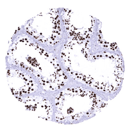
Recombinant Rabbit monoclonal / IgG 1:100 – 1:200 Research Use Only Nuclear Human MSVA-403R BHLHE33 Kidney: A moderate to strong TFE3 staining should be seen in a fraction of glomerular cells and an at least weak TFE3 staining should be seen in a subset of tubular and collecting duct cells. Colon: TFE3 staining should be absent in epithelial cells (some very faint staining can occasionally occur at the surface). TFE3 is a Critical regulator of expression of genes involved in lysosomal function. The Transcription factor E3 (TFE3) protein is a member of the microphthalmia-associated transcription factor (MITF) family of the basic helix-loop-helix leucine zipper family of transcription factors. It is coded by the TFE3 gene at chromosome Xp11.2. In non-starving, resting cells, TFE3 is primarily cytoplasmic. In case of lack of nutrition or energy stress, TFE3 translocates to the nucleus and becomes active. TFE3 target genes play a role in control of metabolic processes in response to nutrient and energy stress and especially include genes implicated in the control of lysosomal functions, such as endocytosis, phagocytosis, and autophagy. As such, TFE3 is involved in metabolic responses and metabolic homeostasis in various tissues. A subset of 1-5% of sporadic renal cell carcinomas are driven by gene fusions that cause upregulation of one of the microphthalmia-associated transcription factor (MiT) family members TFE3, TFEB, or MITF. These fusion driven cancers may make up for more t... Images describing the TFE3 staining pattern in normal tissues obtained by the antibody MSVA-403R are shown in our “ Normal Tissue Gallery ”. Brain Cerebrum Some astrocytic cells show faint TFE3 positivity. Cerebellum Endocrine Tissues Thyroid Weak to moderate TFE3 staining can be seen in follicular cells. Parathyroid Some epithelial cells can show faint TFE3 staining. Adrenal gland Moderate to strong TFE3 staining of cortical cells. Pituitary gland Moderate staining of a variable number of epithelial cells. Respiratory system Respiratory epithelium TFE3 staining of (mainly basal) epithelial cells of the respiratory epithelium and of glandular cells can be seen in some samples. Lung A large subset of pneumocytes and of alveolar macrophages show weak to moderate TFE3 positivity. Gastrointestinal Tract Salivary glands Few glandular cells may show weak TFE3 staining. Esophagus A weak TFE3 staining of squamous epithelial cells can occasionally be seen. Stomach Mucosa epithelial cells are us... Although TFE3 staining is characteristic for a Xp11.2 translocation RCC, TFE3 immunostaining – at variable levels of intensity – can be found in many different tumor types. The TCGA findings on TFE3 RNA expression in different tumor categories have been summarized in the Human Protein Atlas. Xp11.2 fusion carcinoma with strong TFE3 staining of all tumor cells Squamous cell carcinoma with weak TFE3 positivity of large fraction of tumor cells TFE3 negative clear cell carcinoma (2) Cancer tissue gallery No data available at the moment IHC users have different preferences on how the stains should look like. Some prefer high staining intensity of the target stain and even accept some background. Others favor absolute specificity and lighter target stains. Factors that invariably lead to more intense staining include higher concentration of the antibody and visualization tools, longer incubation time, higher temperature during incubation, higher temperature and longer duration of the heat induced epitope retrieval (slide pretreatment). The impact of the pH during slide pretreatment has variable effects and depends on the antibody and the target protein. All images and data shown here and in our image galleries are obtained by the manual protocol described below. Other protocols resulting in equivalent staining are described as well. Manual protocol Freshly cut sections should be used (less than 10 days between cutting and staining). Heat-induced antigen retrieval for 5 minutes in an autoclave at 121°C in pH 7,8 Target ... The prognostic role of variable TFE3 expression levels in RCCs without TFE3 fusions need to be investigated. The diagnostic and prognostic relevance of TFE3 expression in other tumors and in preneoplastic disease needs to be investigated. There are two ways how the specificity of antibodies can be documented for immunohistochemistry on formalin fixed tissues. These are: 1. Comparison with a second independent method for target expression measurement across a large number of different tissue types (orthogonal strategy), and 2. Comparison with one or several independent antibodies for the same target and showing that all positive staining results are also seen with other antibodies for the same target (independent antibody strategy). Orthogonal validation: For proteins such as TFE3 which are expressed in virtually all tissues but restricted to specific cell types and cell compartments, orthogonal validation is not suited. However, the comparison of MSVA-403R immunostaining data with RNA expression data from three independent RNA screening studies, including the Human Protein Atlas (HPA) RNA-seq tissue dataset, the FANTOM5 project, and the Genotype-Tissue Expression (GTEx) project, which are all summarized in the Human Pro...