TYMS (HMV305)
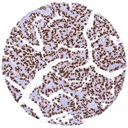
Recombinant Rabbit monoclonal / IgG 1:100 – 1:200 Research Use Only Intracellular Human HMV305 thymidylate synthetase , HST422 , TMS , TS Lymph node: A strongTYMSstaining should be seen in a fraction of lymphocytic cells, especially in germinal centres. Prostate:TYMSstaining should be absent in normal epithelial cells. TYMS is a critical enzyme for thymidine synthesis and folate metabolism. Thymidylate synthase (TS, TYMS) is a 32-35kD enzyme which is coded by the TYMS gene at 18p11.32. TYMS catalyzes the conversion of deoxyuridine monophosphate (dUMP) to deoxythymidine monophosphate (dTMP) which is one of the nucleotides forming the DNA. TYMS is essential for DNA synthesis because it represents the only de novo pathway for production of thymidine and it also is the only enzyme in folate metabolism that can oxidize the 5,10-methylenetetrahydrofolate during one-carbon transfer. Therefore, TYMS is critical for regulating the supply of all 4 DNA precursors for DNA replication. In-vitro studies have shown that upregulation of TYMS is sufficient to transform immortalized mammalian cells to a malignant phenotype. TYMS is an important target for several chemotherapeutic drugs including 5-fluorouracil (5-FU). It has been suggested that tumors with low-levels of TYMS may show to a better response to 5-FU than those with high-level expression. In normal tissues, TYMS expression is u... Images describing the TYMS staining pattern in normal tissues obtained by the antibody HMV305 are shown in our “ Normal Tissue Gallery ”. Brain Cerebrum Negative. Cerebellum Negative. Endocrine Tissues Thyroid Negative. Parathyroid Negative. Adrenal gland Negative. Pituitary gland Negative. Respiratory system Respiratory epithelium Negative. Lung Negative. Gastrointestinal Tract Salivary glands Negative. Esophagus Weak to moderate, predominantly nuclear TYMS staining of a subset of suprabasal squamous epithelial cells. Stomach Weak to moderate, nuclear and cytoplasmic TYMS staining of a fraction of epithelial cells. Duodenum Weak, nuclear and cytoplasmic TYMS staining of a fraction of crypt epithelial cells. Small intestine Weak to moderate, nuclear and cytoplasmic TYMS staining of a fraction of crypt epithelial cells. Appendix Weak to moderate, nuclear and cytoplasmic TYMS staining of a fraction of crypt epithelial cells while staining is strong in many lymphocytic cells. Colon Faint,... TYMS expression is highly variable between individual tumors. At least in a fraction of tumors, high TYMS expression occurs in a broad range of different tumor entities. The TCGA findings on TYMS RNA expression in different tumor categories have been summarized in the Human Protein Atlas. Diffuse large B-cell lymphoma with mostly
intense TYMS staining of tumor cells. Testicular seminoma with TYMS staining
limited in some inflammatory cells. Gastrointestinal stromal tumor (GIST) with strong
TYMS staining of a scattered tumor cells. Cancer tissue gallery No data available at the moment IHC users have different preferences on how the stains should look like. Some prefer high staining intensity of the target stain and even accept some background. Others favor absolute specificity and lighter target stains. Factors that invariably lead to more intense staining include higher concentration of the antibody and visualization tools, longer incubation time, higher temperature during incubation, higher temperature and longer duration of the heat induced epitope retrieval (slide pretreatment). The impact of the pH during slide pretreatment has variable effects and depends on the antibody and the target protein. All images and data shown here and in our image galleries are obtained by the manual protocol described below. Other protocols resulting in equivalent staining are described as well. Manual protocol Freshly cut sections should be used (less than 10 days between cutting and staining). Heat-induced antigen retrieval for 5 minutes in an autoclave at 121°C in pH 7,8 Target ... The diagnostic, prognostic, and predictive role of TYMS expression in tumors and in preneoplastic disease needs to be investigated. There are two ways how the specificity of antibodies can be documented for immunohistochemistry on formalin fixed tissues. These are: 1. Comparison with a second independent method for target expression measurement across a large number of different tissue types (orthogonal strategy), and 2. Comparison with one or several independent antibodies for the same target and showing that all positive staining results are also seen with other antibodies for the same target (independent antibody strategy). Orthogonal validation: For the antibody HMV305 specificity is supported by the good concordance of the immunostaining data with data from three independent RNA screening studies, including the Human Protein Atlas (HPA) RNA-seq tissue dataset, the FANTOM5 project, and the Genotype-Tissue Expression (GTEx) project, which are all summarized the Human Protein Atlas (Tissue expression TYMS) . TYMS positivity by HMV305 is detectable at highest levels in these tissues with highest TYMS RNA expressio...