Villin (MSVA-459R)
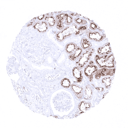
Recombinant Rabbit monoclonal / IgG 1:100 – 1:200 Research Use Only Cell Surface and Cytoplasmic Human MSVA-459R VIL1; Villin-1; Villin1 Colon: A moderate to strong villin staining with predominance of the apical membrane should be seen in all epithelial cells. Liver: A weak to moderate staining of the apical pole of hepatocytes should be seen. Colon: Villin staining should be absent in all non-epithelial cells. Villin is a 92.5 kDa protein encoded by the VIL1 gene located at the human chromosome 2q35. The protein is mainly localized in the microvilli of the brush border of various normal epithelial cell types. As an actin-binding protein, Villin is connected to the actin core bundle of the brush border. Villin has actin modifying functions and is involved in the nucleation, crosslinking, capping and splitting of actin filaments. Amongst others, the actin modifying functions are regulated by calcium and phosphorylation of the villin protein. Functional studies suggest an impact of Villin upregulation on the cytoskeleton architecture, cellular shape and motility as well as cell viability. Villin is a 92.5 kDa protein encoded by the VIL1 gene located at the human chromosome 2q35. The protein is mainly localized in the microvilli of the brush border of various normal epithelial cell types. As an actin-binding protein, Villin is connected to the actin core bundle of the brush border. Villin has actin modifying functions and is involved in the nucleation, crosslinking, capping and splitting of actin filaments. Amongst others, the actin modifying functions are regulated by calcium and phosphorylation of the villin protein. Functional studies suggest an impact of Villin upregulation on the cytoskeleton architecture, cellular shape and motility as well as cell viability. Villin staining pattern in Normal Tissues with antibody MSVA-459R (images are shown in our “Normal Tissue Gallery”) Brain Cerebrum Negative. Cerebellum Negative. Endocrine Tissues Thyroid Negative. Parathyroid Negative. Adrenal gland Negative. Pituitary gland Negative. Respiratory system Respiratory epithelium Negative. Lung Negative. Gastrointestinal Tract Salivary glands Negative. Esophagus Negative. Stomach Weak to moderate villin positivity of surface epithelium. Gradual decrease of staining intensity towards the base of the glands. Duodenum Strong villin positivity in all epithelial cells. Small intestine Strong villin positivity in all epithelial cells. Appendix Strong villin positivity in all epithelial cells. Colon Strong villin positivity in all epithelial cells. Rectum Strong villin positivity in all epithelial cells. Liver Weak to moderate villin staining of luminal membranes of hepatocytes (zonal variability of staining intensity). Gallbladder Negative. Pancreas Strong vill... In carcinomas, villin expression is predominantly cytoplasmic. Villin positivity is most commonly seen in colorectal cancer. Tumors that were described to also show villin expression in a variable fraction of cases include gastric, duodenal and esophageal carcinoma, gastrointestinal neuroendocrine tumors, endometrial carcinoma, hepatocellular carcinoma, pulmonary adenocarcinoma (enteric type) and others. The TCGA findings on Villin RNA expression in different tumor categories have been summarized in the Human Protein Atlas. Moderate to strong villin staining in all cells of a colorectal adenocarcinoma showing apical membrane predominance. Strong villin immunostaining in a muscle-invasive urothelial carcinoma of the urinary bladder Moderate to strong, predominantly apical villin staining in all cells of an endometrioid carcinoma of the ovary. Cancer tissue gallery Villin (MSVA-459R) publication summary Relevant publication: Dum et al. “Villin expression in human tumours: a tissue microarray study on 14,398 tumours ” . Published in Expert Review of Molecular Diagnostics Accepted 18 Jul 2022, Accepted author version posted online: 22 Jul 2022, PMID: 35866621 A total of 12,429 tumors were successfully analyzed from 118 different tumor categories by using the following protocol: Heat-induced antigen retrieval for 5 minutes in an autoclave at 121°C in pH7,8 Target Retrieval Solution buffer. MSVA-459R at a dilution of 1:250 at 37°C for 60 minutes. Visualization of bound antibody by the EnVision Kit (Dako, Agilent). This protocol was also used for all stainings depicted in our tumor and normal tissue galleries. In this study, at least one positive case was seen in 54 (46%) of 118 tumor categories and 36 (31%) tumor categories included at least one case with strong positivity The highest frequencies of villin positivity (and the highest levels of expre... IHC users have different preferences on how the stains should look like. Some prefer high staining intensity of the target stain and even accept some background. Others favor absolute specificity and lighter target stains. Factors that invariably lead to more intense staining include higher concentration of the antibody and visualization tools, longer incubation time, higher temperature during incubation, higher temperature and longer duration of the heat induced epitope retrieval (slide pretreatment). The impact of the pH during slide pretreatment has variable effects and depends on the antibody and the target protein. Accordingly, multiple different protocols can generate identical staining results. All images and data shown here and in our image galleries are obtained by the manual protocol described below. Other protocols resulting in equivalent staining are described as well. -Manual protocol Freshly cut sections should be used (less than 10 days between cutting and staining). Hea... The clinical/prognostic significance of villin expression in individual tumor types is unknown. Specificity of MSVA-459R is documented by strong positive staining in cell types that are well documented to express villin such as epithelial cells of the colorectum, appendix, small intestine, duodenum, pancreatic excretion ducts and in proximal tubuli of the kidney as well as absence of staining in all tissues known to not express villin including tissues notorious for non-specific IHC background such squamous epithelium or distal tubuli of the kidney. The validity of the normal tissue staining patterns is also confirmed by a comparison of MSVA-459R staining with staining results obtained by a second independent, commercially available antibody (RTU IVD). Antibody comparison: MSVA-459R vs another commercial anti-Villin antibody RTU (IVD) MSVA-459R – Epididymis MSVA-459R – Liver MSVA-459R – Pancreas MSVA-459R – Stomach RTU (IVD) – Epididymis RTU (IVD) – Liver RTU (IVD) – Pancreas RTU (IVD) – Stomach Normal tissue gallery