Vimentin (MSVA-458R)
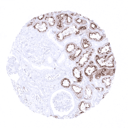
Recombinant Rabbit monoclonal / IgG 1:100 – 1:200 Research Use Only Cytoplasmic Human MSVA-458R VIM Liver: Kupffer cells must show a strong staining and sinusoidal endothelial cells should show an at least a weak staining. Colon: Endothelial and muscle cells of large vessels and stromal cells must show a strong Vimentin staining while dispersed intraepithelial T-cells must show an at least moderate intensity staining. Vimentin is a 57 kDa protein coded by the VIM gene at 10p13. It is the first intermediate filament protein to be expressed during cell differentiation. All primitive cell types express vimentin but in most non-mesenchymal cells it is later replaced by other intermediate filament proteins. In non-vascular smooth muscle cells and in striated muscle, vimentin is often replaced by desmin, but vimentin remains the major intermediate filament seen in non-muscle cells. It can be re-expressed in muscle cells in case of injury and regeneration. Vimentin is required for stabilizing the position of the organelles in the cytosol. Vimentin provides cells with resilience and is considered the cytoskeletal component responsible for maintaining cell integrity. Transgenic mice that lack vimentin are viable although they show various developmental defects and delayed wound healing. Vimentin is the major cytoskeletal component of mesenchymal cells and is thus used as a marker of mesenchymal cell origin and of epithelial-to-mesenchymal transition (EMT) during cancer progression. Vimentin expression induces a transformation of cells to an elongated, flat, mesenchymal shape. Vimentin is a 57 kDa protein coded by the VIM gene at 10p13. It is the first intermediate filament protein to be expressed during cell differentiation. All primitive cell types express vimentin but in most non-mesenchymal cells it is later replaced by other intermediate filament proteins. In non-vascular smooth muscle cells and in striated muscle, vimentin is often replaced by desmin, but vimentin remains the major intermediate filament seen in non-muscle cells. It can be re-expressed in muscle cells in case of injury and regeneration. Vimentin is required for stabilizing the position of the organelles in the cytosol. Vimentin provides cells with resilience and is considered the cytoskeletal component responsible for maintaining cell integrity. Transgenic mice that lack vimentin are viable although they show various developmental defects and delayed wound healing. Vimentin is the major cytoskeletal component of mesenchymal cells and is thus used as a marker of mesenchymal cell origin a... Vimentin immunostaining is found in various mesenchymal cells including fat cells, fibroblasts, endothelial cells, macrophages, melanocytes, Langerhans cells, Schwann cells, glial cells, lymphocytes, mesothelium, ovarian granulosa cells, Sertoli and Leydig cells of the testis. Vimentin is usually absent in skeletal and heart muscle, but regularly seen in vascular smooth muscle. In non-vascular smooth muscle vimentin expression is normally low or absent, but can be upregulated in case of regeneration. Vimentin is also regularly found in several specialized epithelia, such as the Bowman capsule of the kidney, fallopian tube, endometrium, endocervix (weak), thyroid gland, adrenal gland (cortex and medulla), and pancreas (basolateral portion of acinar cells) as well as in myoepithelial cells of the breast, salivary and sweat glands. Vimentin can occasionally also be seen in some normal appearing prostate acinar cells, principal cells of the epididymis, scattered respiratory epithelial cell... Vimentin is present in many different neoplasms but is particularly expressed in those originated from mesenchymal cells. Sarcomas e.g., fibrosarcoma, angiosarcoma, and leio- and rhabdomyosarcoma, sarcoma NOS, as well as lymphomas, malignant melanoma and schwannoma, are virtually always vimentin positive. Mesoderm derived carcinomas like renal cell carcinoma, adrenal cortical carcinoma and adenocarcinomas from endometrium and ovary usually express vimentin. Also thyroid carcinomas are vimentin positive. Any poorly differentiated or sarcomatoid carcinoma may express some vimentin. The TCGA findings on Vimentin RNA expression in different tumor categories have been summarized in the Human Protein Atlas. Papillary renal cell carcinoma with strong baso-lateratal vimentin staining of all tumor cells Prostatic adenocarcinoma (Gleason 3+3=6) showing strong vimentin staining limited to the tumor stroma Strong vimentin imunostaining of the tumor stroma in a muscle-invasive urothelial carcinoma ... No data available at the moment IHC users have different preferences on how the stains should look like. Some prefer high staining intensity of the target stain and even accept some background. Others favor absolute specificity and lighter target stains. Factors that invariably lead to more intense staining include higher concentration of the antibody and visualization tools, longer incubation time, higher temperature during incubation, higher temperature and longer duration of the heat induced epitope retrieval (slide pretreatment). The impact of the pH during slide pretreatment has variable effects and depends on the antibody and the target protein. Accordingly, multiple different protocols can generate identical staining results. All images and data shown here and in our image galleries are obtained by the manual protocol described below. Other protocols resulting in equivalent staining are described as well. Manual protocol Freshly cut sections should be used (less than 10 days between cutting and staining). Heat... Vimentin is a canonical marker for epithelial-mesenchymal transformation. As such it can be used in multicolor imaging for delineating and studying cells undergoing EMT. Specificity of MSVA-458R is documented by strong positive staining in all mesenchymal cell types that are well documented to express vimentin and absence of staining in skeletal and heart muscle as well as in these epithelial tissues known to not express vimentin such as for example colon epithelium and hepatocytes. Normal tissue gallery